
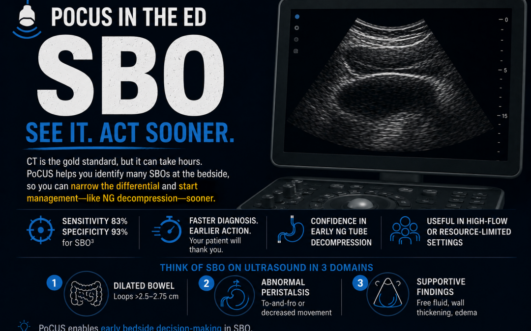
Small bowel obstruction (SBO) is a common ED presentation, and these patients are often in significant pain with persistent nausea and vomiting. While CT remains the gold standard to confirm the diagnosis, identify the transition point, detect complications such as bowel ischemia, and guide surgical management, it can often take hours to obtain. During that time, patients continue to suffer while awaiting definitive imaging.
PoCUS allows you to identify many SBOs at the bedside, helping narrow the differential diagnosis, accelerate management, and identify patients who may benefit from early nasogastric decompression while awaiting CT. In settings where CT is delayed or unavailable, PoCUS also offers substantially greater diagnostic accuracy than plain radiography.¹˒²
Why PoCUS Matters
- Sensitivity 83% and specificity 93% for PoCUS in the identification of SBO3
- Faster diagnosis and cognitive offloading
- Confidence in earlier empiric management with NG tube before confirmatory CT (your patient will thank you…after the discomfort of inserting the NG)
- Useful in resource-limited or high-flow ED settings
Mental Model: Think of SBO on Ultrasound in 3 Domains:
- Dilated bowel
- Abnormal peristalsis
- Supportive findings (free fluid, wall changes)
How to Perform the Scan
Set up
-
-
- Probe: Curvilinear
- Depth: 10–15 cm
- Patient: Supine
-
Technique
-
-
- Start in RLQ
- Use graded compression to displace bowel gas and assess for bowel compressibility
- Sweep all quadrants using a “Zamboni technique” (“Lawnmower” for you non-Canucks; see Fig 1)
- Scan in two planes (i.e., short- and long-axis probe orientation)
-
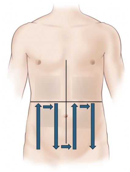
Fig 1. Zamboni technique when scanning for SBO. Arrows indicate systematic movement of ultrasound probe across the abdomen
Findings Suggestive of SBO
Dilated bowel diameter:
- ≥2.5 cm(traditional threshold for diagnosis of SBO on PoCUS; Sn 85.2%, Sp 59.1%)4
- ≥2.75 cm(more balanced diagnostic accuracy; Sn 71.8%, Sp 67.1%)4
- <1.7 cmrules out SBO with 100% sensitivity4
- Pearl: Bowel diameter is measured from inner-wall to inner-wall
- Pearl: Measure bowel diameter in the short-axis viewto avoid inaccuracies by the cylinder tangent effect (See Fig 2)
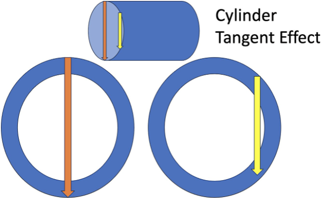
Fig 2. Cylinder tangent effect. Ultrasound artifact where a cylindrical organ (e.g., dilated bowel, aorta) appears artificially small or falsely narrow due to the imaging plane being off-centre, resulting in a tangent or side-view image rather than the maximum diameter
Peristalsis
- Early findings: to-and-fro and/or swirling movement of bowel content (see below)
- Late findings: decreased/absent movement of bowel content.
- Pearl: How do I know if decreased/absent movement of bowel content is a late finding of SBO vs. a normal finding in someone who may just have slow moving bowel normally? Answer: look for the presence of supportive findings(see below) and consider the clinical context as well!
Supportive findings:
- “Tanga” sign(named for its similar appearance to a bikini bottom): a triangle-shaped fluid collection located between dilated loops of bowel (see Fig 53).
- Bowel wall thickening >3mm (see Fig 6)
- Pearl: measure thickness at the bowel wall closest to your probe (i.e., the near-field of the screen) to avoid artificial enlargement via posterior acoustic enhancement artifact.
- “Piano key” sign: bowel wall edema in the setting of SBO can accentuate the appearance of plicae circularis into the bowel lumen (see Fig 74).
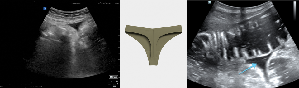
Fig 3. Tanga sign. Triangle-shaped free-fluid between dilated loops of bowel. Indicative of high-grade and advanced obstruction.
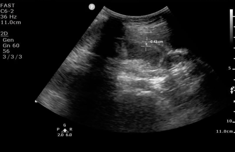
Fig 4. Example of bowel wall thickening. This example measures at 4.3mm
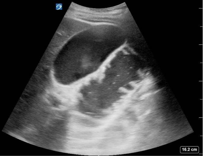
Fig 5. Piano key sign. Bowel wall edema emphasizes the appearance of small bowel plicae circularis (blue arrow), giving the appearance of hyperechoic piano keys projecting into the hypoechoic bowel lumen.
Troubleshooting
- Use gentle compression of the abdominal wall with your ultrasound probe to displace bowel gas that may be obscuring your image
- Have your patient bend their hips and knees to help relax the abdominal wall
Pitfalls
- Ileus can mimic SBO
- No single finding is definitive
- Cannot reliably predict need for surgery
- Can be very challenging to identify transition point
- Negative scan does not exclude early SBO
Bottom Line
- Look for dilated loops (>2.5–2.75 cm)
- Assess peristalsis
- Use supportive findings
- PoCUS enables early bedside decision-making in SBO.
References
- Kidmas, A. T., Ekedigwe, J. E., Sule, A. Z., & Pam, S. D. (2005). A review of the radiological diagnosis of small bowel obstruction using various imaging modalities. Nigerian Postgraduate Medical Journal, 12(1), 33–36.
- Pourmand, A., Dimbil, U., Drake, A., & Shokoohi, H. (2018). The accuracy of point-of-care ultrasound in detecting small bowel obstruction in the emergency department. Emergency Medicine International, 2018, 3684081. https://doi.org/10.1155/2018/3684081
- Shokoohi, H., Mayes, K. D., Peksa, G. D., Loesche, M. A., Becker, B. A., Boniface, K. S., Lahham, S., Jang, T. B., Secko, M., & Gottlieb, M. (2023). Multi-center analysis of point-of-care ultrasound for small bowel obstruction: A systematic review and individual patient-level meta-analysis. American Journal of Emergency Medicine, 70, 144–150. https://doi.org/10.1016/j.ajem.2023.05.039
- Shokoohi, H., Al Jalbout, N., Peksa, G. D., Mayes, K. D., Becker, B. A., Boniface, K. S., Lahham, S., Secko, M., Chavoshzadeh, M., Jang, T. B., & Gottlieb, M. (2024). Optimal bowel diameter thresholds for diagnosing small bowel obstruction and surgical intervention with point-of-care ultrasound. American Journal of Emergency Medicine, 84, 1–6. https://doi.org/10.1016/j.ajem.2024.07.019