
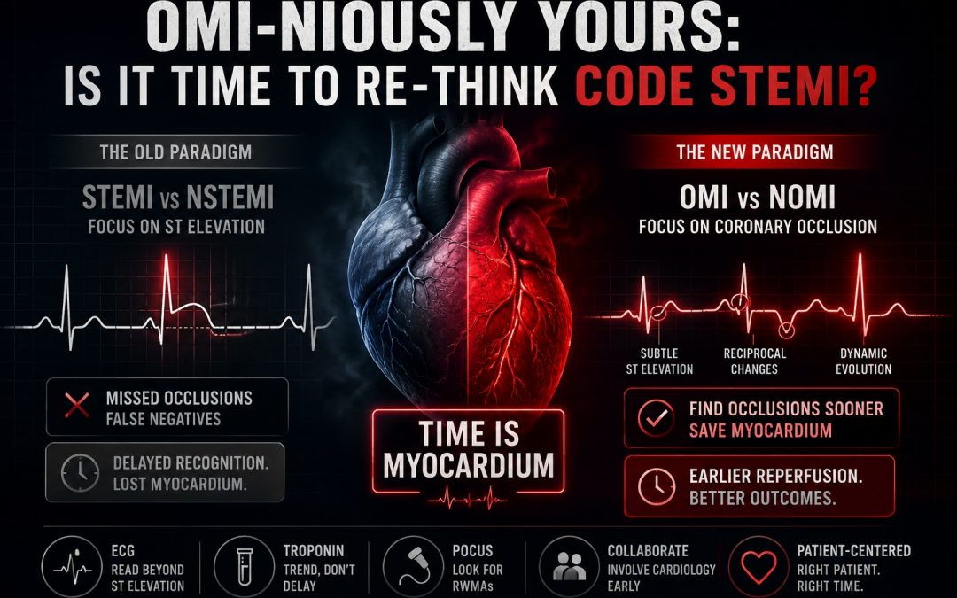
Cardiovascular disease remains one of the leading causes of death worldwide, accounting for approximately 32% of deaths globally.¹ Acute myocardial infarction (MI) accounts for more than 50,000 deaths annually in Canada.² As emergency physicians, we all recognize the surge of urgency when a “Code STEMI” is called overhead. But it may be time to reconsider whether this familiar paradigm serves our patients as well as we think it does.
How We Got Here: The Birth of STEMI
To understand the limitations of the STEMI paradigm, it helps to understand how we got here. For much of the 20th century, myocardial infarctions were retrospectively classified according to whether patients developed Q waves on their ECG.³ With little available to alter the course of an evolving infarction, the distinction between “Q-wave” and “non-Q-wave” MI was primarily descriptive.
Then came reperfusion.
Streptokinase was first discovered in 1933, and decades later thrombolytic therapy transformed the treatment of acute MI.⁴ By the late 20th century, large randomized trials demonstrated that fibrinolysis could reduce mortality in acute MI, particularly when given early and in patients presenting with ST-segment elevation or bundle branch block. The landmark 1994 Fibrinolytic Therapy Trialists’ meta-analysis pooled data from approximately 58,600 patients across nine randomized trials and confirmed a significant mortality benefit from fibrinolytic therapy.⁴
Suddenly, waiting for Q waves to appear was no longer good enough. If opening an acutely occluded coronary artery could save myocardium and lives, clinicians needed a way to identify these patients early.
The ECG became that tool.
Over time, ST-segment elevation became the primary surrogate for identifying patients who required immediate reperfusion. Current criteria define significant ST elevation as new elevation at the J point in at least two contiguous leads, generally ≥1 mm, with different thresholds in V2-V3 based on age and sex.⁵
This gave rise to the STEMI/NSTEMI paradigm that has dominated acute coronary syndrome care for decades. Patients meeting STEMI criteria receive immediate reperfusion, while those classified as NSTEMI generally follow a less emergent pathway.
There is just one problem: ST elevation and acute coronary occlusion are not the same thing.
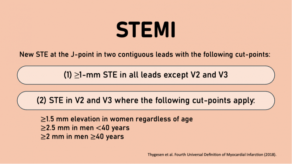
Figure 1. STEMI Criteria based on the Fourth Universal Definition of Myocardial Infarction
This paradigm shift reinforced the concept that “time is myocardium” and catalyzed decades of research into angiography, PCI, and adjunctive medical therapy.⁶ It also led to interdisciplinary QI efforts to reduce door-to-balloon times, including rapid ED ECG acquisition, ED physician-initiated cath lab activation, paramedic bypass of the ED, and close collaboration with interventional cardiology.⁶ Thus, our beloved Code STEMI was born.
Cracks in the Code: Where the STEMI Paradigm Is Failing Us
1. The Paradigm is Unquestioned
Some of the limitations of the STEMI paradigm can be traced back to the trials that established the benefit of fibrinolysis. But how did these studies determine whether patients actually had an occluded coronary artery in the first place?
They didn’t.
Routine angiography was not performed, and MI was diagnosed using the biomarkers available at the time, such as CK-MB, rather than contemporary high-sensitivity troponin assays.⁶ The FTT authors themselves cautioned against rigid interpretation of the ECG when deciding who should receive reperfusion therapy, noting that “certain cases require experienced interpretation of the ECG before withholding reperfusion therapy.”⁶
Yet, in 2024, a meta-analysis by de Alencar et al. found that despite decades of reliance on STEMI criteria, only three studies had evaluated their diagnostic accuracy for detecting angiographically confirmed acute coronary occlusion. The pooled sensitivity was just 43.6%.⁶
In other words, the ECG criteria we have built an entire reperfusion system around may miss more than half of acute coronary occlusions.
2. The False-Negative Paradox
If the STEMI paradigm perfectly identified acute coronary occlusion, patients with an occluded culprit artery would reliably meet STEMI criteria. In reality, they don’t.
Like any diagnostic test, STEMI criteria produce true positives, true negatives, false positives, and false negatives. But our terminology tends to hide the false negatives. When a patient has an acute coronary occlusion but does not meet STEMI criteria, we don’t call it a “false-negative STEMI.” We call it an NSTEMI.⁴
That semantic distinction has real consequences.
In 2017, Khan et al. published a meta-analysis of seven studies including approximately 40,000 patients diagnosed with NSTEMI.⁴˒⁷ Among these patients, 25.5% had an occluded culprit artery on angiography.⁴˒⁷ Patients with an occluded culprit artery also had higher rates of major adverse cardiovascular events and a 1.5-fold higher relative risk of mortality compared with NSTEMI patients without an occluded artery.⁴˒⁷
In 2021, Meyers et al. prospectively studied 467 ED patients with suspected ACS.⁸ Among patients with confirmed occlusion MI, only 62% met STEMI criteria.⁸ This means 38% of patients with an OMI did not meet STEMI criteria.
The difference in treatment was striking. Patients with OMI who met STEMI criteria had a median time from arrival to catheterization of 41 minutes. Those with OMI who did not meet STEMI criteria waited a median of 437 minutes.⁸
We all know that time is myocardium. Yet our dichotomized STEMI/NSTEMI paradigm can place a patient with an acutely occluded coronary artery onto a pathway where emergent reperfusion is no longer the default.
3. False-Positive STEMIs
The problem also exists in the opposite direction. Approximately 15–35% of cath lab activations have been reported to result in angiography without a culprit coronary lesion.⁹
This does not necessarily mean the activation was inappropriate. Some degree of overtriage is inevitable, and arguably desirable, in a system designed to rapidly identify a time-sensitive emergency. However, unnecessary cath lab activation is not benign. Angiography carries risks including vascular and bleeding complications, coronary dissection or perforation, kidney injury related to contrast exposure, and resource utilization.¹⁰
Taken together, these limitations expose a fundamental problem with the STEMI paradigm: ST-segment elevation is an imperfect surrogate for the pathology we actually care about, an acutely occluded coronary artery causing ongoing myocardial infarction.
As physicist and philosopher Thomas Kuhn wrote, “Failure of existing rules is the prelude to a search for new ones.”⁶
Enter OMI: A Paradigm Shift
In 2018, two emergency physicians, Dr. Stephen Smith and Dr. Pendell Meyers, published the OMI Manifesto in collaboration with EMCrit.³ In it, they proposed the term occlusion myocardial infarction (OMI) to describe an acute coronary occlusion or near-occlusion causing ongoing myocardial infarction.¹⁰ Conversely, non-occlusion myocardial infarction (NOMI) describes an MI without an acute culprit occlusion, or where sufficient collateral circulation maintains perfusion despite the occlusion.¹⁰
Rather than relying on ST elevation as the primary trigger for emergent reperfusion, the OMI/NOMI paradigm focuses on the underlying pathology we actually care about: is there an acutely occluded coronary artery causing ongoing myocardial infarction that requires emergent reperfusion? Importantly, this framework also makes the false negatives of traditional STEMI criteria visible.
In 2020, Aslanger et al. published the DIFOCCULT study, examining whether ECG findings beyond traditional STEMI criteria could identify patients with acute coronary occlusion who had initially been classified as NSTEMI.¹¹ Two cardiologists, blinded to angiographic outcomes, reviewed ECGs from patients with STEMI, NSTEMI, and control groups.¹¹
The results were striking: 28.2% of patients initially classified as NSTEMI were reclassified as having an ECG suspicious for acute coronary occlusion.¹¹ These patients had higher rates of acute coronary occlusion and myocardial injury. Moreover, patients with NSTEMI and an occluded culprit artery had in-hospital and long-term mortality that more closely resembled the STEMI group than NSTEMI patients without an occlusion.¹¹
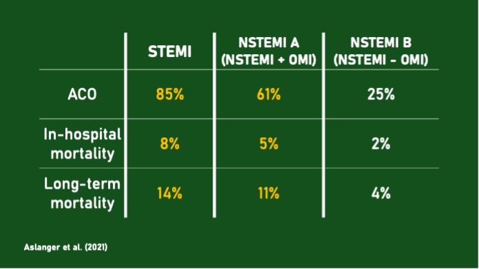
Similarly, in 2021, Meyers et al. conducted a retrospective case-control study using data from patients with ACS who underwent cardiac catheterization over a one-year period.¹² Among 808 patients, 265 met the study definition of OMI. Of these, only 108 met STEMI criteria, giving traditional STEMI criteria a sensitivity of just 41% for OMI.¹²
When investigators blinded to the clinical outcomes interpreted the ECGs using additional features of acute coronary occlusion beyond traditional STEMI criteria, they identified 38 additional OMIs that had been missed by STEMI criteria.¹² Importantly, these ECG findings were present a median of approximately three hours before angiography was actually performed, suggesting an opportunity for substantially earlier recognition and reperfusion.¹²
So what were we missing on the ECG?
As shown in Figure 3, STEMI-negative OMI was rarely electrocardiographically silent. Subtle ST elevation that did not meet STEMI criteria was present in 83%, while reciprocal ST depression or T-wave inversion was present in 82%.¹² Several other findings were present in approximately half of cases, and only 4% of OMIs had none of the identified ECG features.¹²
In other words, many of these occlusions weren’t invisible on the ECG. They simply didn’t meet STEMI criteria.
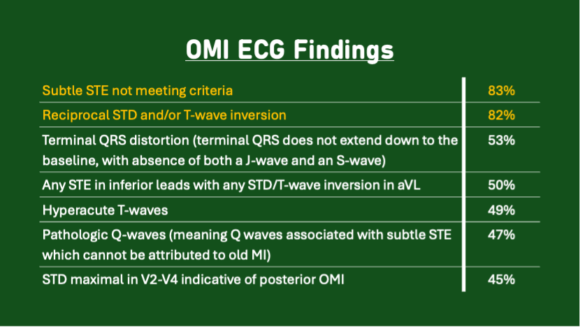
Figure 3. ECG findings from study by Myers et al. (2021)
Spotting the Occlusion: How to Diagnose OMIs
ECG remains the cornerstone of the OMI paradigm because it is rapid, widely available, and non-invasive. However, OMI is ultimately a clinical diagnosis, and other diagnostic tools, including serial ECGs, troponin, and point-of-care ultrasound (POCUS), can provide important additional information when clinical suspicion remains high despite a nondiagnostic initial ECG.
1. ECG
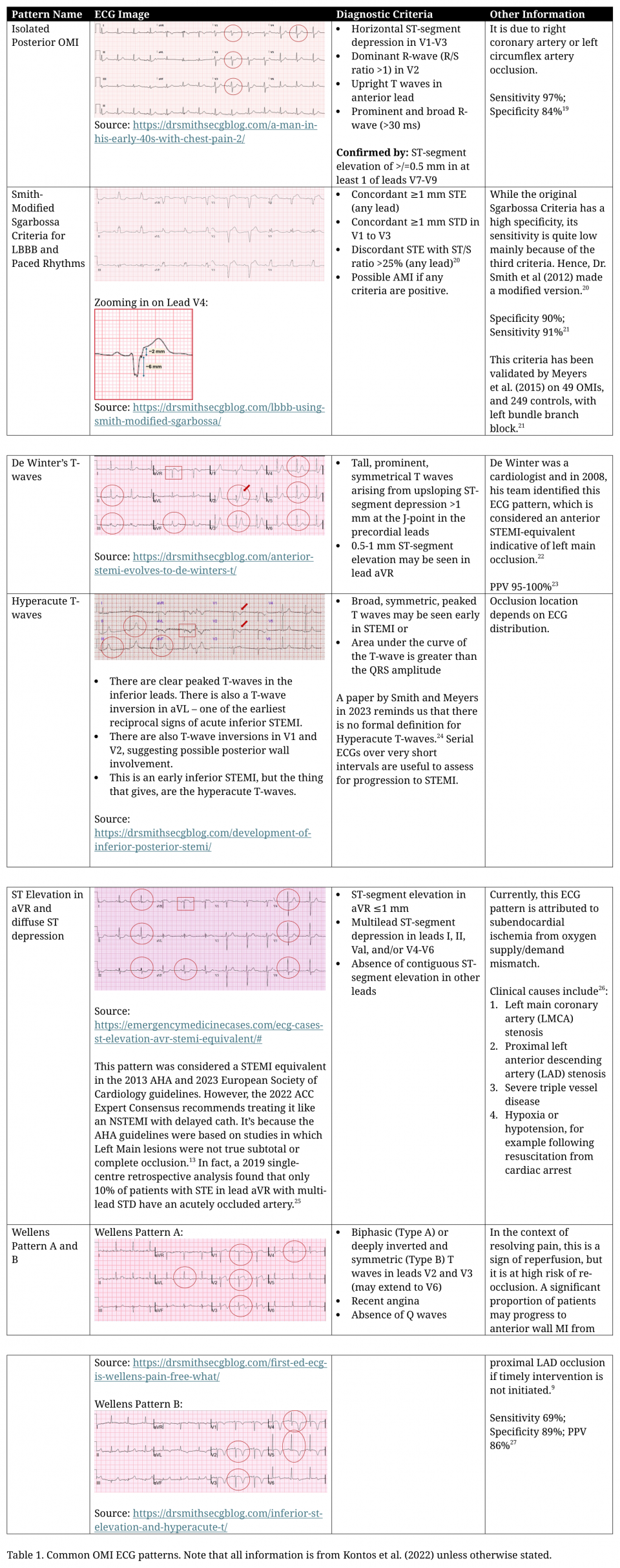
More than 20 ECG patterns associated with OMI have been described, although many have not yet been prospectively validated. Table 1 highlights several of the better-established patterns, including those recognized in clinical guidelines and the 2022 American College of Cardiology Expert Consensus Decision Pathway on the Evaluation and Disposition of Acute Chest Pain in the Emergency Department.
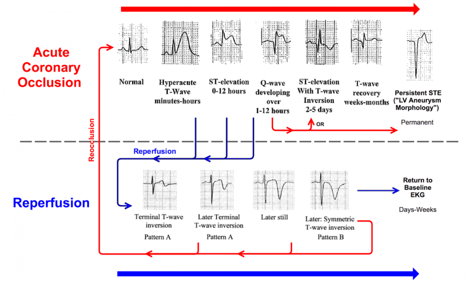
Figure 4. Dynamic ECG changes of acute coronary occlusion. Source: https://hqmeded-ecg.blogspot.com/
One of the most important concepts in OMI is that coronary occlusion is a dynamic process, and so is the ECG. The same patient’s tracing can look dramatically different depending on when it is obtained.¹³ Early in an occlusion, hyperacute T waves may appear before diagnostic ST elevation develops. With persistent occlusion, ST elevation and eventually Q waves may develop.
The ECG can also evolve when spontaneous reperfusion occurs. As blood flow is restored, ST segments may normalize and terminal T-wave inversion can develop. With continued reperfusion, these T waves may become deeper and more symmetric. Importantly, an ECG obtained during this phase may no longer meet STEMI criteria, despite the patient having recently experienced an acute coronary occlusion.
This is why a single ECG is only a snapshot. Serial ECGs, interpreted in the context of the patient’s symptoms and compared with previous tracings whenever possible, can reveal an evolving occlusion that a single ECG may miss.
For a deeper dive into specific OMI ECG patterns, the following resources provide excellent reviews:
- Ricci et al (2024): https://www.sciencedirect.com/science/article/pii/S0196064424012502
- OMI Guide: https://omiguide.org/
2. Troponin
Troponin reflects myocardial injury rather than what is happening in the coronary artery at that exact moment. In patients presenting early after symptom onset, even high-sensitivity troponin may initially be normal or only minimally elevated. Therefore, a negative initial troponin should not reassure you when the history and ECG remain concerning for OMI.¹³˒¹⁴
In STEMI-negative patients with persistent symptoms, however, troponin can provide important additional evidence of ongoing myocardial injury. Serial measurements are particularly useful, as a significant rise can support the diagnosis when the initial ECG is nondiagnostic. However, waiting for troponin elevation can delay recognition and reperfusion of OMI.¹⁴
The key is to use troponin as part of the overall clinical picture rather than as a gatekeeper for reperfusion. When there is compelling evidence of acute coronary occlusion, treatment should not be delayed while waiting for biomarkers.
3. PoCUS
POCUS can be a powerful adjunct in suspected OMI, particularly when the ECG is nondiagnostic. Acute myocardial ischemia can produce a regional wall motion abnormality (RWMA), which may be visible on ultrasound before significant biomarker elevation.¹⁵ A new RWMA corresponding to a coronary territory can therefore increase suspicion for OMI in an otherwise equivocal presentation.
However, a normal echocardiogram does not exclude early AMI. POCUS is operator dependent, and subtle wall motion abnormalities can be difficult to identify. For this reason, it is better used to “rule in” than “rule out” OMI.¹⁵ Multiple cardiac views should be obtained rather than relying on the parasternal short-axis view alone.
POCUS can also identify alternative diagnoses and mechanical complications of MI, including acute mitral regurgitation from papillary muscle dysfunction or rupture, ventricular septal rupture, pericardial effusion, and features concerning for free-wall rupture.¹⁶ One important limitation is that acute and chronic RWMAs may look similar, making comparison with a previous echocardiogram particularly valuable when available.
So, what does the evidence show? A 2023 retrospective review found that emergency physician-performed POCUS for RWMA had a sensitivity of 94% but a specificity of only 35%, and identified abnormalities in 87% of patients with angiographically confirmed OMI.¹⁴ Another single-centre study found a substantially shorter time to revascularization among patients with OMI when a RWMA was identified: 432 minutes compared with 2,158 minutes when no RWMA was identified.¹⁷
POCUS therefore shows promise as an adjunct in suspected OMI, particularly when the ECG does not provide a definitive answer. But like troponin, it should complement rather than replace careful clinical and ECG assessment.
A Practical Approach: How to Think OMI
According to the 2022 ACC Expert Consensus Decision Pathway on the Evaluation and Disposition of Acute Chest Pain in the Emergency Department, patients who meet STEMI criteria, have a STEMI-equivalent ECG, or are clinically unstable with high suspicion for ACS should undergo emergent evaluation for revascularization.¹⁸ Other high-risk ischemic patterns, including Wellens syndrome, also warrant prompt Cardiology involvement but do not necessarily trigger immediate cath lab activation.¹⁸
In practice, however, things are less clear-cut. The OMI/NOMI framework has not yet been fully incorporated into Cardiology guidelines, while our existing STEMI systems are supported by decades of evidence and highly developed regional protocols. OMI recognition is also more nuanced than traditional STEMI criteria, raising legitimate concerns about prematurely expanding cath lab activation pathways and the resource implications that could follow.
Unlike a Code Stroke, which prioritizes rapid diagnostic evaluation despite considerable diagnostic uncertainty, a Code STEMI mobilizes an entire cath lab team, particularly after hours, to deliver a definitive intervention for presumed acute coronary occlusion. Any expanded activation strategy therefore needs to maintain an acceptable positive predictive value, something the evolving OMI literature has not yet consistently demonstrated.
For clear STEMIs, activate a Code STEMI. When the ECG does not meet STEMI criteria but there is concern for OMI, the next step will depend on your local system. In many centres, early direct discussion with Cardiology may be more appropriate than automatically activating the cath lab.
When communicating concern for a possible OMI, clarity is key. Rather than simply saying “the patient doesn’t meet STEMI criteria,” communicate the pathology you are worried about: acute coronary occlusion with ongoing myocardial ischemia. Integrate the patient’s symptoms, dynamic or subtle ECG findings, hemodynamics, serial troponin measurements, and POCUS findings when available. These additional findings should strengthen the overall clinical assessment rather than act as prerequisites for consultation or reperfusion.
Antithrombotic therapy should follow contemporary ACS guidelines and local protocols, with Cardiology input when the diagnosis or anticipated revascularization strategy is uncertain. Similarly, fibrinolysis should be reserved for patients who meet established indications when timely primary PCI is not available, rather than being administered solely on the basis of a suspected OMI pattern.
For now, cath lab activation should continue to follow institutional and regional pathways. But when the ECG falls short of STEMI criteria and the rest of the clinical picture suggests an acute coronary occlusion, “NSTEMI” should not be the end of the diagnostic thought process. Recognize the possibility of OMI, involve Cardiology early, and advocate for timely reperfusion when the evidence points toward an acutely occluded artery.
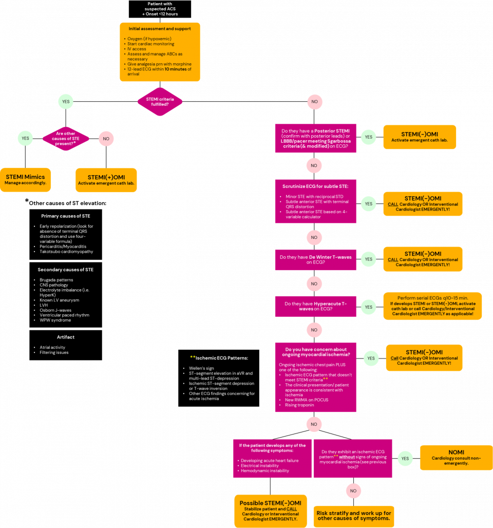
Figure 5. Suggested practical approach to OMI identification and cardiology consultation.
Where Do We Go From Here?
Moving from a STEMI/NSTEMI framework toward OMI/NOMI will require more than recognizing a few additional ECG patterns. It will require prospective validation, evidence that earlier OMI recognition improves patient-centred outcomes, and collaboration between emergency medicine, cardiology, paramedicine, and regional systems of care.
Education will also remain key. OMI recognition relies on interpreting the ECG as a whole, including proportionality, reciprocal changes, subtle abnormalities, and dynamic changes over time. Like any ECG skill, this improves with practice, continuing medical education, case review, and discussion with colleagues and consultants.
Artificial intelligence may also have a role. The Queen of Hearts model, incorporated into PMCardio, was developed specifically to identify ECG evidence of acute coronary occlusion beyond traditional STEMI criteria.⁹ The technology is increasingly being evaluated across different clinical settings, with promising but variable results. AI may eventually provide another set of “eyes” on the ECG, particularly for subtle occlusions, but it should currently be viewed as an adjunct rather than a replacement for clinical judgment, ECG expertise, and appropriate Cardiology consultation.
Key Takeaways
#1: STEMI criteria are a tool, not the disease.
The pathology we ultimately care about is an acutely occluded coronary artery causing ongoing myocardial infarction. STEMI criteria identify many of these patients quickly and reliably, but they do not identify all of them.
#2: Read the whole ECG, not just the ST segments.
Look for subtle and proportional ST changes, hyperacute T waves, reciprocal changes, and other high-risk patterns. Compare with previous ECGs and repeat the ECG when symptoms change. Remember that coronary occlusion is dynamic, with ECG findings evolving during occlusion, reperfusion, and re-occlusion.
#3: A STEMI-negative ECG should not end the conversation when the clinical picture is concerning.
Integrate the ECG with the patient’s symptoms, hemodynamics, serial troponins, and POCUS when appropriate. If you remain concerned about acute coronary occlusion, involve Cardiology early and advocate for your patient.
The STEMI paradigm transformed the treatment of acute myocardial infarction and has saved countless lives. OMI does not ask us to abandon that progress. It asks us to recognize where the existing framework may fall short and to look beyond the millimetres when the patient in front of us is telling us something is wrong.
Time is myocardium. Our job is to recognize whose myocardium is still at risk.
References
- Cardiovascular diseases (CVDs) [Internet]. World Health Organization; 2025 [cited 2026 Apr 26]. Available from: https://www.who.int/en/news-room/fact-sheets/detail/cardiovascular-diseases-(cvds)
- Public Health Agency of Canada. Surveillance of heart diseases and conditions [Internet]. Government of Canada. 2017 [cited 2026 Apr 26]. Available from: https://www.canada.ca/en/public-health/services/diseases/heart-health/heart-diseases-conditions/surveillance-heart-diseases-conditions.html
- Meyers P, Scott W, Stephen S. The OMI Manifesto [Internet]. Dr. Smith’s ECG Blog. 2018 [cited 2026 Apr 26]. Available from: https://drsmithsecgblog.com/the-omi-manifesto/
- Aslanger EK, Meyers HP, Smith SW. Time for a new paradigm shift in myocardial infarction. The Anatolian Journal of Cardiology [Internet]. 2021 Mar 4 [cited 2026 Apr 26];25(3):156–62. Available from: https://pmc.ncbi.nlm.nih.gov/articles/PMC8114732/
- Thygesen K, Alpert JS, Jaffe AS, Chaitman BR, Bax JJ, Morrow DA, et al. Fourth Universal Definition of Myocardial Infarction (2018). Circulation [Internet]. 2018 Aug 24 [cited 2026 Apr 26];138(20). Available from: https://www.ahajournals.org/doi/10.1161/CIR.0000000000000617
- McLaren J, de Alencar JN, Aslanger EK, Meyers HP, Smith SW. From ST-Segment Elevation MI to Occlusion MI: The New Paradigm Shift in Acute Myocardial Infarction. JACC: Advances [Internet]. 2024 Nov [cited 2026 Apr 26];3(11):101314. Available from: https://www.sciencedirect.com/science/article/pii/S2772963X24005933
- Rezaie S. Rebellion21: Time to Evolve – Redefining Coronary Ischemia – The OMI/NOMI Paradigm via Tarlan Hedayati, MD [Internet]. REBEL EM – Emergency Medicine Blog. 2021 [cited 2026 Apr 26]. Available from: https://rebelem.com/rebellion21-time-to-evolve-redefining-coronary-ischemia-the-omi-nomi-paradigm-via-tarlan-hedayati-md/
- Murali S. OMI-NOMI: Time for a Paradigm Shift [Internet]. REBEL EM – Emergency Medicine Blog. 2021 [cited 2026 Apr 26]. Available from: https://rebelem.com/omi-nomi-time-for-a-paradigm-shift/
- Ayyad M, Albandak M, Gala D, Alqeeq B, Baniowda M, Pally J, et al. Reevaluating STEMI: The Utility of the Occlusive Myocardial Infarction Classification to Enhance Management of Acute Coronary Syndromes. Current Cardiology Reports [Internet]. 2025 Mar 27 [cited 2026 Apr 24];27(1). Available from: https://pmc.ncbi.nlm.nih.gov/articles/PMC11950105/#Sec2
- Buttner R, Cadogan M. OMI: Replacing the STEMI misnomer [Internet]. Life in the Fast Lane. 2021 [cited 2026 Apr 26]. Available from: https://litfl.com/omi-replacing-the-stemi-misnomer/
- Alblaihed L. The DIFOCCULT Trial: Time to Change from STEMI/NSTEMI to OMI/NOMI? [Internet]. REBEL EM – Emergency Medicine Blog. 2020 [cited 2026 Apr 22]. Available from: https://rebelem.com/the-difoccult-trial-time-to-change-from-stemi-nstemi-to-omi-nomi/
- Pendell Meyers H, Bracey A, Lee D, Lichtenheld A, Li WJ, Singer DD, et al. Accuracy of OMI ECG findings versus STEMI criteria for diagnosis of acute coronary occlusion myocardial infarction. IJC Heart & Vasculature [Internet]. 2021 Apr [cited 2026 Apr 26];33(33):100767. Available from: https://pmc.ncbi.nlm.nih.gov/articles/PMC8065286/
- Miranda DF, Lobo AS, Walsh B, Sandoval Y, Smith SW. New Insights Into the Use of the 12-Lead Electrocardiogram for Diagnosing Acute Myocardial Infarction in the Emergency Department. Canadian Journal of Cardiology [Internet]. 2018 Feb [cited 2026 Apr 26];34(2):132–45. Available from: https://pubmed.ncbi.nlm.nih.gov/29407007/
- Al-Zaiti S, Martin-Gill C, Zègre-Hemsey J, Bouzid Z, Faramand Z, Alrawashdeh M, et al. Machine Learning for the ECG Diagnosis and Risk Stratification of Occlusion Myocardial Infarction at First Medical Contact. Research Square (Research Square) [Internet]. 2023 Jan 30 [cited 2026 Apr 26]; Available from: https://pubmed.ncbi.nlm.nih.gov/36778371/
- Bracey A, Massey L, Pellet AC, Thode HC, Holman TR, Singer AJ, et al. FOCUS may detect wall motion abnormalities in patients with ACS. The American journal of emergency medicine [Internet]. 2023 Jul 1 [cited 2026 Apr 26];69:17–22. Available from: https://pubmed.ncbi.nlm.nih.gov/37037160/
- McLaren J. ECG Cases 41 – STEMI, Occlusion MI Complications [Internet]. Emergency Medicine Cases. 2023 [cited 2026 Apr 24]. Available from: https://emergencymedicinecases.com/stemi-occlusion-mi-complications/
- Xu C, Melendez A, Nguyen T, Ellenberg J, Anand A, Delgado J, et al. Point-of-care ultrasound may expedite diagnosis and revascularization of occult occlusive myocardial infarction. The American Journal of Emergency Medicine [Internet]. 2022 Aug 1 [cited 2026 Apr 26];58:186–91. Available from: https://pubmed.ncbi.nlm.nih.gov/35700615/
- Kontos MC, de Lemos JA, Deitelzweig SB, Diercks DB, Gore MO, Hess EP, et al. 2022 ACC Expert Consensus Decision Pathway on the Evaluation and Disposition of Acute Chest Pain in the Emergency Department. Journal of the American College of Cardiology [Internet]. 2022 Oct [cited 2026 Apr 26];80(20). Available from: https://www.jacc.org/doi/10.1016/j.jacc.2022.08.750
- EOF van Gorselen, FWA Verheugt, BTJ Meursing, AJM Oude Ophuis. Posterior myocardial infarction: the dark side of the moon. Netherlands Heart Journal [Internet]. 2007 Jan [cited 2026 Apr 26];15(1):16. Available from: https://pmc.ncbi.nlm.nih.gov/articles/PMC1847720/
- Cadogan M, Buttner R. Sgarbossa Criteria [Internet]. Life in the Fast Lane. 2019 [cited 2026 Apr 28]. Available from: https://litfl.com/sgarbossa-criteria-ecg-library/
- Macfarlane PW. New ECG Criteria for Acute Myocardial Infarction in Patients With Left Bundle Branch Block [Internet]. J Am Heart Assoc; 2024 [cited 2026 Apr 26]. Available from: https://pmc.ncbi.nlm.nih.gov/articles/PMC7660728/#jah35237-bib-0004
- Buttner R, Burns E. De Winter T Wave [Internet]. Life in the Fast Lane. 2021 [cited 2026 Apr 26]. Available from: https://litfl.com/de-winter-t-wave/
- Morris NP, Body R. The De Winter ECG pattern: morphology and accuracy for diagnosing acute coronary occlusion: systematic review. European Journal of Emergency Medicine [Internet]. 2017 Aug [cited 2026 Apr 26];24(4):236–42. Available from: https://pubmed.ncbi.nlm.nih.gov/28362646/
- Smith SW, Meyers HP. Hyperacute T-waves Can Be a Useful Sign of Occlusion Myocardial Infarction if Appropriately Defined. Annals of Emergency Medicine [Internet]. 2023 Mar 1 [cited 2026 Apr 26];82(2). Available from: https://www.annemergmed.com/article/S0196-0644(23)00025-2/abstract
- Harhash AA, Huang JJ, Reddy S, Natarajan B, Balakrishnan M, Shetty R, et al. aVR ST Segment Elevation: Acute STEMI or Not? Incidence of an Acute Coronary Occlusion. The American Journal of Medicine [Internet]. 2019 May [cited 2026 Apr 26];132(5):622–30. Available from: https://www.amjmed.com/article/S0002-9343(19)30049-X/fulltext
- Buttner R, Burns E. ST Elevation in aVR [Internet]. Life in the Fast Lane. 2020 [cited 2026 Apr 26]. Available from: https://litfl.com/st-elevation-in-avr/
- Das D, Almajed NS. Wellens syndrome. Canadian Medical Association Journal [Internet]. 2016 Jan 11 [cited 2020 Aug 28];188(7):529–9. Available from: https://pmc.ncbi.nlm.nih.gov/articles/PMC4835285/