
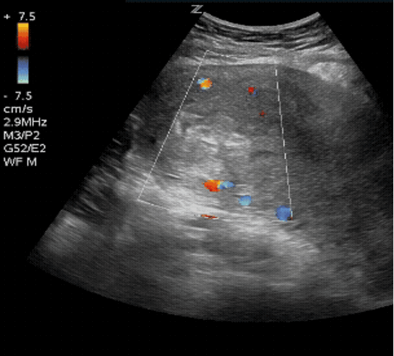
It’s 2:30 AM. A 29-year-old woman presents ten days after a medical abortion. She’s febrile, tachycardic, and complaining of worsening pelvic pain. She has a small amount of vaginal bleeding, and her abdomen is soft. Her β-hCG is still positive, which is expected. Your community hospital has no overnight ultrasound and the nearest Gynecologist is two hours away. Is this simply pain following her completed abortion…or is retained products of conception (RPOC) after an incomplete abortion driving sepsis?
You perform a bedside PoCUS which reveals a thickened endometrium containing heterogeneous echogenic material with internal vascularity on Doppler.
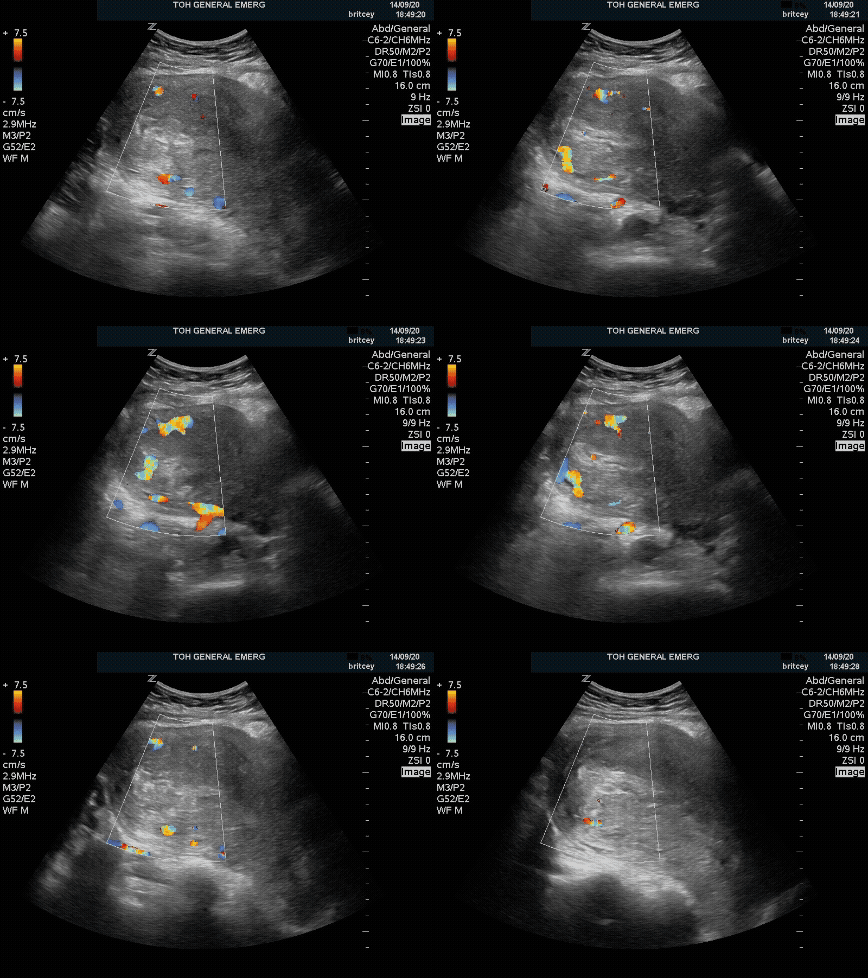
Within minutes you’ve started antibiotics, called Gynecology, and arranged urgent transfer for suspected retained products of conception (RPOC).
While radiology-performed ultrasound remains the diagnostic standard, PoCUS can help rapidly identify concerning findings at the bedside, especially in settings where comprehensive imaging may be delayed or unavailable.
This PoCUS Pearl reviews how to identify RPOC with PoCUS, what findings matter most, and the major pitfalls Emergency Physicians should keep in mind.
Why you Should Start Scanning for RPOC
Case integration: In the patient above, identifying vascular retained tissue changed management from observation to immediate antibiotics, gynecology consultation and transfer.
Patients with RPOC commonly present with:
- Vaginal bleeding after miscarriage, abortion, or delivery
- Pelvic pain or cramping
- Persistent positive β-hCG
- Fever or infectious symptoms (less common, but concerning)
Importantly, physical exam and laboratory findings are often nonspecific. PoCUS can:
- Help rapidly identify concerning intrauterine pathology
- Support earlier OB/GYN consultation
- Increase confidence in initiating management
- Be particularly useful in resource-limited or after-hours settings where comprehensive ultrasound is delayed
A 2025 retrospective study by Boivin et al. found that for identifying RPOC in patients with a history concerning for RPOC, transabdominal PoCUS had: [1]
- Sensitivity: 79%
- Specificity: 93.8%
The takeaway? PoCUS appears particularly useful as a “rule-in” tool when classic findings are present.
What are Retained Products of Conception?
RPOC refers to residual intrauterine tissue remaining after:
- Miscarriage
- Medical or surgical abortion
- Vaginal delivery
- Cesarean delivery
- The retained material may include:
- Placental tissue
- Decidual tissue
- Blood products
- Fetal tissue
This retained tissue can become vascular and continue bleeding or become infected and lead to endometritis or septic abortion. Additionally, long-term, retained tissue can cause uterine adhesions potentially adversely affecting fertility.
How to Perform the Scan
Probe
- Curvilinear probe
- Endocavitary (transvaginal) probe if available and within your own scope/comfort level
Preset
- Obstetric or pelvic preset
Patient Position/Preparation
- Supine
- A full bladder improves transabdominal visualization, but an empty bladder improves a transvaginal view
Scanning Technique
Transabdominal Scan
- Place the probe suprapubically in the sagittal orientation with the marker toward the patient’s head
- Identify the bladder first
- Visualize the uterus posterior to the bladder
- Sweep through the uterus from side to side
- Rotate to transverse orientation and sweep from cervix to fundus
- Carefully assess the endometrial cavity
What Should You Focus On?
Three major things:
- Endometrial thickness
- Echogenic intrauterine material
- Internal vascularity
Key PoCUS Findings
1. Thickened Endometrium
A thickened endometrial stripe is one of the most important findings in RPOC.vSuggested thresholds vary in the literature:
- 7 mm: concerning
- 10 mm: commonly used threshold
- 15 mm: more specific but less sensitive
Boivin et al. defined RPOC as:
- Heterogeneous intrauterine material
- Measuring >10 mm
Pearl: A thin endometrium makes significant RPOC less likely.
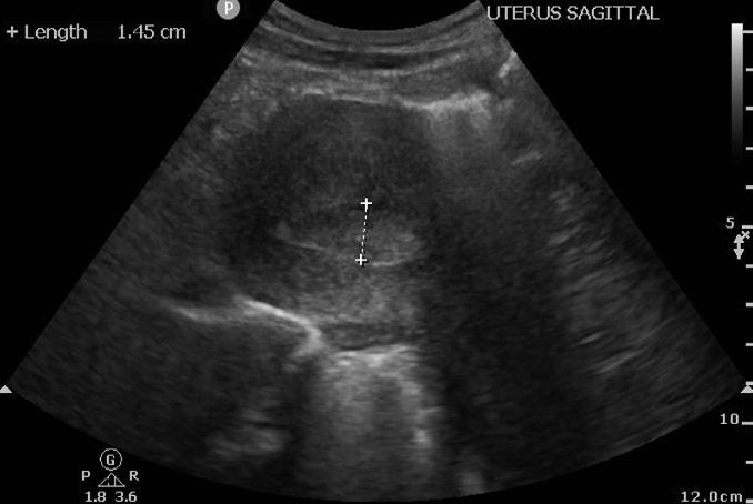
Fig 1. Example of thickened endometrium on transabdominal view (adapted from Boivin et al., 2025)
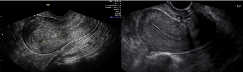
Fig 2. Example of thickened endometrium (left) vs normal uterus (right) on transvaginal view (adapted from Sonosession, 2024 and Dobaria, 2025, respectively)
2. Echogenic or Heterogeneous Material Within the Endometrial Cavity
Look for:
- Echogenic tissue
- Heterogeneous material
- Irregular intrauterine contents
This can appear as:
- Clumpy echogenic debris
- Mixed echogenicity tissue
- Thickened heterogeneous endometrium
Pearl: Blood clot and RPOC can appear similar on grayscale imaging alone, so turn on color Doppler to assess for vascularity and differentiate between the two.
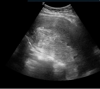
Fig 3. Echogenic material seen within thickened endometrial cavity
3. Internal Vascularity (Very Important)
Color Doppler demonstrating vascular flow within endometrial material is highly suggestive of RPOC.
Why?
- Blood clots are generally avascular.
- Retained placental tissue often remains vascular.
Look for:
- Internal color flow within echogenic material
- Increased endometrial vascularity
Pearl: Internal vascularity is one of the most specific ultrasound findings for RPOC; however, absence of vascularity does NOT exclude RPOC. Some retained products may be avascular (sometimes referred to as hematometra). If clinical suspicion is high, escalate to Radiology ultrasound or Gynecology consultation.
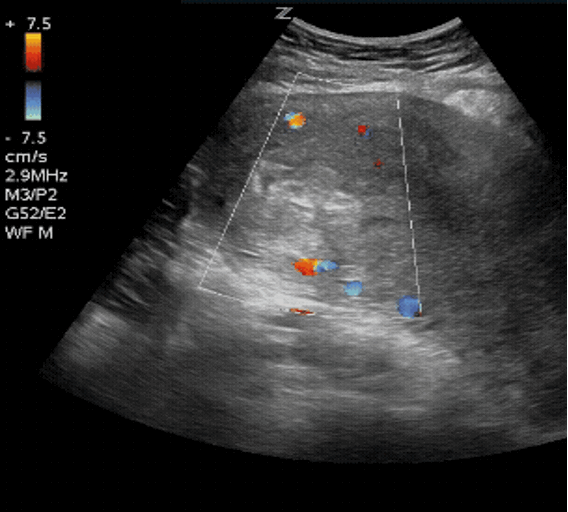
Fig 4. Increased internal vascularity within endometrial material
Example Mental Model
Think of RPOC using 3 ultrasound domains:
- Thickened endometrium
- Echogenic intrauterine material
- Internal vascularity
The more findings present, the more concerning the scan becomes.
Troubleshooting Pearls
Trouble Visualizing the Uterus?
- Ensure adequate bladder filling
- Apply gentle suprapubic pressure to displace bowel gas
- Decrease depth once the uterus is identified
- Sweep slowly through the uterus in two planes
Color Doppler Not Showing Flow?
- Reduce depth
- Ensure the Doppler box is centered over the endometrium
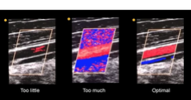
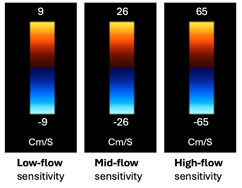
- Remember that very low-flow states may still appear avascular, so decrease your color scale
- If you don’t see any color flow, first increase the color gain until speckle appears, then back it off slightly (see below
- If flow still isn’t visible, lower the color scale to detect slower blood flow (see below).
Pitfalls
1. Early Pregnancy Can Mimic RPOC
Early pregnancies can produce:
- Thickened endometrium
- Heterogeneous uterine contents
- Variable sonographic appearances
Takeaway: Use caution diagnosing RPOC purely from transabdominal PoCUS in early pregnancy. When uncertain, don’t hesitate to order a comprehensive ultrasound.
2. Blood Clot vs RPOC
Both can appear echogenic.
Color Doppler helps differentiate:
- Blood clot → usually avascular
- RPOC → often vascular
3. Fibroids and Other Uterine Pathology
Fibroids may:
- Distort the endometrium
- Create heterogeneous echogenicity
- Mimic intrauterine masses
Always interpret findings within the clinical context.
4. A Negative Scan Does Not Fully Exclude RPOC
PoCUS has imperfect sensitivity. If clinical suspicion remains high:
- Obtain comprehensive ultrasound
- Involve OB/GYN
- Ensure close follow-up
Bottom Line
- Think about RPOC in patients with post-pregnancy bleeding or pelvic pain
- Focus on:
- Endometrial thickness
- Echogenic intrauterine material
- Internal vascularity
- Doppler flow within endometrial material is highly suggestive of RPOC
- Use caution in early pregnancy where mimics are common
- PoCUS can accelerate diagnosis and consultation, particularly when comprehensive imaging is delayed
Recognizing these findings at the bedside won’t replace comprehensive ultrasound, but it may be the reason antibiotics are started an hour earlier, the transfer is arranged before radiology arrives in the morning, or the septic patient gets to the operating room before she deteriorates.
References