
Unstable pelvic fractures have high mortality rates, particularly with patients who are hemodynamically unstable, due to difficulty in achieving hemostasis and other associated injuries. At present, there is no standard guideline that has been published and universally accepted in the management of pelvic trauma. These patients should have integrated management between ED physicians, trauma surgeons, orthopedic surgeons and interventional radiologists. In the words of Scott Weingart, “If you don’t have an institutional protocol, you are going to fail… this needs to be worked out before the crashing patient comes in!”
Objectives:
- Review important concepts in the mechanical stability of pelvic fractures
- Discuss ED management and key interventions in controlling hemorrhage
- Outline an algorithm to manage pelvic trauma in the ED
Anatomy Review
- The pelvis is made up of the innominate bones (pubis, ischium, ilium), the sacrum, and coccyx
- Because the pelvis is structured as a ring, a break at one point is often associated with disruption at a second point
- The stability of the ring depends on the rigidity of the bones and the integrity of the ligaments that bind the three segments together across the symphysis pubis and the sacroiliac joints
- The venous system is the most common source of significant pelvic bleeding in addition to bleeding from cancellous bone
- Arterial bleeds comprise about 10% of pelvic bleeding
Classifications for Hip Fracture
- The Young Burgess Classification is most commonly used to emphasize the mechanism of injury by vector and severity
- Lateral Compression: primarily caused by a lateral force, but may involve anterior or posterior vector components
- Antero-Posterior Compression: a normal symphysis diastasis is less than 0.5cm, but we can accept a diastasis up to 2.5cm
- An unstable “open book” symphysis pubis measures >2.5cm
- Vertical shear injuries: usually occur with an axial load to the hemipelvis
- The Tile Classification, unlike Young Burgess, outlines the biomechanical stability of the pelvis and not the mechanism
- Below is my interpretation on how to understand the YB and Tile classifications in terms of mechanical stability, and how these may influence management of the patient in the ED
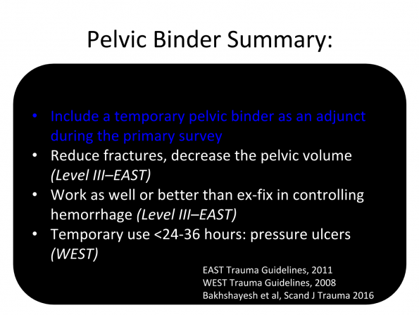
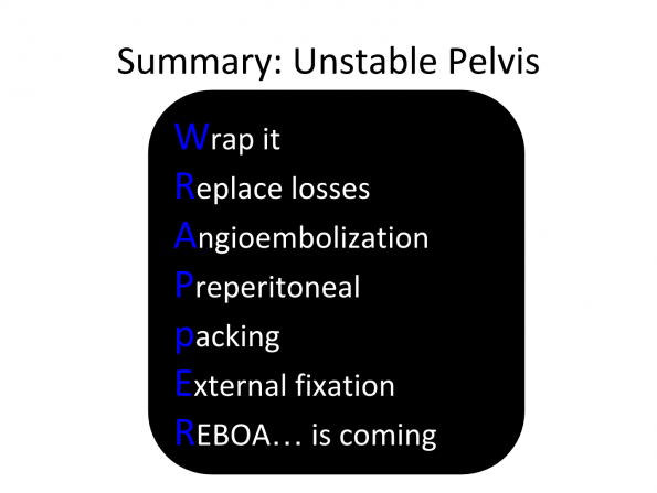
Stabilizing an Unstable Pelvis in the ED
- Placement of an external fixator reduces the fracture and can decrease the pelvic volume by 10-20%
- Historically, these were being used early in management, but did not help to control hemorrhage and were ultimately delaying hemorrhage control
- An external fixator can be applied by Orthopaedic Surgery once the patient is hemodynamically stable, or in conjunction with pre-peritoneal packing in the OR
- Temporary pelvic binders are now favoured to fixation methods in the ED because they are much faster to apply
- They should be placed over the greater trochanters and pubic symphysis, not over the abdomen
Diagnostic Tools and interventions in management of pelvic fractures:
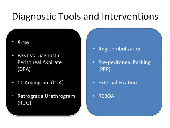
X-rays:
- Pelvic X-ray should be incorporated into a trauma primary survey, unless there is complete absence of pain on physical exam without significant mechanism, distracting injury, intoxication, or decreased level of consciousness
- Pelvic fracture pattern itself does not predict mortality, hemorrhage, or the need for angiography–unstable fractures have been associated with increased risk of bleeding, but significant bleeds can occur with mechanically stable fractures as well
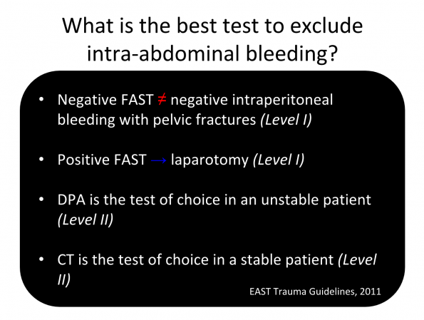
What is the best test to exclude intra-abdominal bleeding in pelvic fractures?
- There is some controversy about whether FAST, Diagnostic peritoneal lavage (DPL), or aspirate (DPA) should be used
- Although the specificity of the FAST in patients who have pelvic fractures is reasonable as an initial screening tool (87-100%), the sensitivity in mechanically unstable Tile B or C pelvic fractures is low (with reports of around 75% but as low as 25% sensitivity)
- Pelvic fractures are a special consideration for DPL because there is a possibility that a large retroperitoneal hematoma may dissect into the anterior abdominal wall, but DPL has been shown to have a high rate of false positives in patients with pelvic fractures
- An aspirate, without the lavage seems to offer similar sensitivity with a lower rate of false negatives.
- Because of the low sensitivity of FAST with pelvic fracture, CTA abdo/pelvis is recommended in patients who are hemodynamically stable
CT Angiogram
- CT contrast extravasation is highly predictive of arterial injury that will require angiographic embolization
- CT has been a reliable indicator of arterial hemorrhage with Sensitivity of 60-90% and Specificity of 85-98% reported
- Absence of contrast extravasation on CT does not always exclude active hemorrhage as bleeding can be intermittent (Level II – EAST)
Retrograde Urethrograms in Pelvic Trauma
- Pelvic fractures are also associated with urethral and bladder injuries in 7-25% of cases
- Clinical signs such as perineal or scrotal swelling, blood from the urethra or the presence of a high riding prostate may suggest presence of a urethral injury
- Retrograde urethrograms should be performed before placement of a foley catheter if there are concerns about urethral injury
- However, if RUG is required, it should be performed after the CTA (Level III-EAST) as there is higher rate of indeterminate or false negative CT scans if RUG is performed prior to CTA
Arterial Embolization
- Selective vs non-selective (bilateral internal iliac artery) embolization can be performed to address arterial bleeding, depending on the stability of the patient and vessel that is bleeding
- Small vessel bleeds are embolized with coils, polyvinyl alcohol, or a glue, which are permanent
- If a larger vascular territory is involved, a temporary gelfoam slurry can be used to provide immediate hemostasis, with less chance of long term ischemia to the area
EAST Trauma Guidelines for Angioembolization in Pelvic Trauma:
- Active arterial extravasation on CTA (Level I)
- Hemodynamic instability without extra-pelvic blood loss (Level I)
- Consideration for stable patient >60 years old with major pelvic disruption (Level II)
- Consider repeat angioembolization if there is ongoing bleeding (Level II)
- Absence of contrast extravasation does not exclude active hemorrhage (Level II)
- Large hematoma >500cm3 (Level III)
- Bilateral non-selective is safe (Level III)
- Male potency is not affected (Level III)
Preperitoneal Packing
- The immediate surgical option for patients is PPP (also called retro-peritoneal packing)
- PPP is a relatively newer method of packing the retroperitoneum without the need for a laparotomy
- PPP is useful for hemodynamically unstable patients with hemorrhage in the pelvis
- Angioembolization can be performed after PPP when the patient is more hemodynamically stable
- PPP can also be performed after failed embolization as a salvage mechanism
- The OR is always the safest place for an unstable pelvic trauma patient as preperitoneal packing + exploratory laparotomy can be done simultaneously, in addition to external fixation of the pelvis
- Studies have shown that PPP in the OR can be done faster than Angioemolization, and that mortality is worse when patients require operative management after failed angioembolization
REBOA: Resuscitative Endovascular Balloon Occlusion of the Aorta
-
- REBOA is available in some EDs and is gaining traction as a bridge until definitive hemorrhage control can be obtained in the OR
- The concept of aortic occlusion in the setting of non-compressive thoracic hemorrhage from the abdomen or pelvis is not new, and has been done in the OR for many years
- Physiologically, occluding the aorta during hemorrhagic shock results in:
- Increased cardiac output
- Increased coronary blood flow
- Increased MAP and perfusion to the brain
- Similar in concept to cross-clamping the aorta (in zone 1), a REBOA catheter could be placed in the infrarenal (zone 3) position using a Seldinger technique
- In effect, this would be similar to placing an arterial line in order to occlude the aorta and prevent blood from further entering a massive unstable pelvic bleed
- The most support for REBOA is in patients who are hypotensive with a systolic BP between 60-80 and have a zone 3 injury
An Algorithm for Managing Pelvic Trauma in the ED
Stable Patient:
- If the patient is hemodynamically stable and the FAST is negative, get a CTA to determine whether there is active contrast extravasation (blush)
- If there is contrast extravasation, angioembolization is indicated
- If no contrast extravasation is seen, but your patient is older than 60 and you have a high suspicion that there is bleeding, angioembolization is worth consideration
Unstable Patient:
- If your patient is hemodynamically unstable, assess where your patient is bleeding from (thoracic, abdominal or pelvic) and manage these life threats appropriately
- Perform a FAST as part of your primary exam
- If the FAST is positive, go to the OR – this is the safest place for an unstable patient as you can pack the pelvis, put on an external fixator if needed, do a laparatomy, and then go to the angio suite afterwards
- If your FAST is negative, organize angioembolization if it can be arranged urgently (30 mins to 1 hour)
- Realistically there can be a clinical gray zone with patients who are somewhat responsive to transfusion and may tolerate a CT scan but are unstable. Keep in mind that all of the literature and guidelines would confirm that unstable patients do not go to the CT scanner, they go to angio or the OR.
- Zone 3 REBOA, if available, is appropriate for hemodynamically unstable patients with negative FAST, but requiring hemodynamic stabilization and hemorrhage control in order to survive transport to the OR
References
- Scott Weingart. Severe Pelvic Trauma. EMCrit Blog. Published on April 30, 2012. Accessed on Sept 12 2017. Available at [https://emcrit.org/emcrit/severe-pelvic-trauma/].
- Cullinane et al. Eastern Association for the Surgery of Trauma Practice Management Guidelines for Hemorrhage in Pelvic Fracture—Update and Systematic Review. The Journal of Trauma: Injury, Infection, and Critical Care, 2011.
- DeAngelis et al. Use of the trauma pelvic orthotic device (T-POD) for provisional stabilisation of anterior–posterior compression type pelvic fractures: A cadaveric study. Injury, 2008.
- Knops et al. Comparison of Three Different Pelvic Circumferential Compression Devices: A Biomechanical Cadaver Study. The Journal of Bone and Joint Surgery-American, 2011.
- Prasarn et al. Comparison of external fixation versus the trauma pelvic orthotic device on unstable pelvic injuries. Journal of Trauma and Acute Care Surgery, 2012.
- Prasarn et al. Comparison of circumferential pelvic sheeting versus the T-POD on unstable pelvic injuries: A cadaveric study of stability. Injury, 2013.
- Nunn et al. Immediate application of improvised pelvic binder as first step in extended resuscitation from life-threatening hypovolaemic shock in conscious patients with unstable pelvic injuries. Injury. 2007.
- Tan et al. Effect of a new pelvic stabilizer (T-POD®) on reduction of pelvic volume and haemodynamic stability in unstable pelvic fractures. Injury, 2010.
- Davis et al. Western Trauma Association Critical Decisions in Trauma: Management of Pelvic Fracture With Hemodynamic Instability. J Trauma: Injury, Infection, and Critical Care, 2008.
- Croce et al. Emergent Pelvic Fixation in Patients with Exsanguinating Pelvic Fractures. Journal of the American College of Surgeons, 2007.
- Ghaemmaghami et al. Effects of early use of external pelvic compression on transfusion requirements and mortality in pelvic fractures. The American Journal of Surgery, 2007.
- Pizanis et al. Emergency stabilization of the pelvic ring: Clinical comparison between three different techniques. Injury, 2013.
- Fu et al. Pelvic circumferential compression devices benefit patients with pelvic fractures who need transfers. American Journal of Emergency Medicine, 2013.
- Bakhshayesh et al. Effectiveness of non invasive external pelvic compression: a systematic review of the literature. Scandinavian Journal of Trauma, Resuscitation and Emergency Medicine, 2016.
- Yoon et al. Pelvic Arterial Hemorrhage in Patients with Pelvic Fractures: Detection with Contrast-enhanced CT. RadioGraphics, 2004.
- Miller et al. External fixation or arteriogram in bleeding pelvic fracture: initial therapy guided by markers of arterial hemorrhage. J Trauma, 2003.
- Hagiwara et al. Predictors of death in patiens with life-threatening pelvic hemorrhage after successful transcatheter arterial embolization. J Trauma, 2003.
- Cerva et al. Detection of bleeding in patients with major pelvic fracture: value of contrast-enhanced CT. Am J Roentgenol, 1996.
- Stephen et al. Early detection of arterial bleeding in acute pelvic trauma. J Trauma, 1999.
- Pereira et al. Dynamic helical computed tomography scan accurately detects hemorrhage in patients with pelvic fracture. Surgery, 2000.
- Netto et al. Retrograde urethrocystophgraphy impairs computed tomography diagnosis of pelvic arterial hemorrhage in the presence of a lower urologic tract injury. J Am Coll Surg 2008.
- Thorson et al. Operating room or angiography suite for hemodynamically unstable pelvic fractures? The Journal of Trauma: Injury, Infection, and Critical Care,2012.
- Li et al. Retroperitoneal packing or angioembolization for haemorrhage control of pelvic fractures—Quasi-randomized clinical trial of 56 haemodynamically unstable patients with Injury Severity Score ≥33. Injury, 2016.
- Biffl et al. The role of REBOA in the control of exsanguinating torso hemorrhage. Journal of Trauma and Acute Care Surgery, 2015.
- Moore et al. Implementation of resuscitative endovascular balloon occlusion of the aorta as an alternative to resuscitative thoracotomy for noncompressible truncal hemorrhage. Journal of Trauma and Acute Care Surgery, 2015.
- Coccolini et al. Pelvic trauma: WSES classification and guidelines. World Journal of Emergency Surgery, 2017.