
Acute valvular emergencies are uncommon, but when they present they can rapidly become life-threatening. Patients often arrive with undifferentiated respiratory failure, pulmonary edema, hypotension, or cardiogenic shock, and the initial diagnosis may not be obvious. Unfortunately, many of the reflexive interventions we rely on in emergency medicine, such as aggressive fluid administration, early intubation, or indiscriminate vasopressor use, can worsen hemodynamics if the underlying physiology is not recognized.
Unlike many other causes of shock, successful management of acute valvular disease depends less on memorizing the specific lesion and more on understanding its hemodynamic consequences. Is the ventricle struggling to eject blood through a fixed obstruction? Or is blood leaking backwards through an incompetent valve? The answer determines whether your priorities should be preserving preload, reducing afterload, supporting contractility, or controlling heart rate.
Although formal echocardiography remains the diagnostic gold standard, point-of-care ultrasound can rapidly identify structural abnormalities and, more importantly, raise suspicion for a valve emergency early in the patient’s resuscitation. Throughout this post, we’ll review the physiology behind the major acute valvular lesions, discuss how POCUS fits into the diagnostic approach, and highlight practical management strategies for the emergency department.
For a deeper in-depth dive into PoCUS for valvular assessment, read here for more.
Aortic Stenosis
For a deeper dive into the acute recognition and management of aortic stenosis, read on here.
Aortic stenosis (AS) is a preload-dependent state characterized by fixed left ventricular outflow obstruction and high afterload, often leading to left ventricular hypertrophy and increased left atrial pressures.4,5
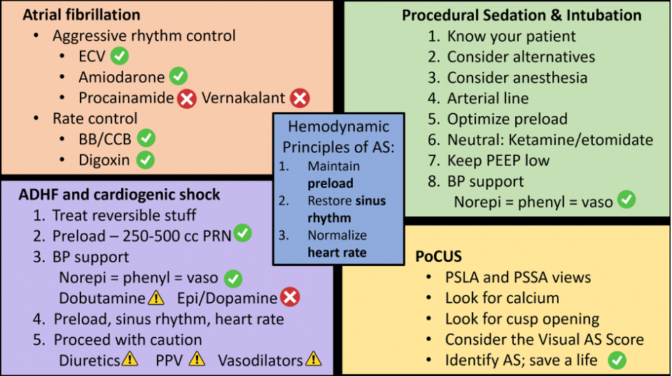
Adequate preload is required to maintain cardiac output due to the noncompliant left ventricle. Hypotension can be catastrophic in patients with severe AS.4 One of the most common mistakes in severe AS is treating hypotension the same way we would septic or distributive shock. Because cardiac output is limited by a fixed obstruction at the aortic valve, patients often cannot compensate with an increase in stroke volume. Even relatively small reductions in preload, whether from diuresis, vasodilation, positive pressure ventilation, or induction medications, can precipitate profound hypotension. Maintaining coronary perfusion pressure is therefore a major priority during resuscitation.
Management principles include cautious fluid administration (if not volume overloaded) and avoidance of aggressive vasodilation.4,6
Vasopressor choice remains debated; norepinephrine, phenylephrine, and vasopressin are all reasonable first-line options, with no clear evidence of superiority.7,8
ED Rules for Aortic Stenosis:
- Hypotension = catastrophe
- Use cautious fluids
- Avoid aggressive vasodilation
- Norepinephrine ≈ Phenylephrine ≈ Vasopressin
Mitral Regurgitation
Mitral regurgitation (MR) results in retrograde blood flow from the left ventricle into the left atrium during systole. Chronic MR allows compensatory remodeling with left ventricular dilation and eccentric hypertrophy.9,10
In acute MR, however, there is sudden regurgitation into a noncompliant left atrium, leading to rapid increases in left atrial and pulmonary venous pressures. Patients can rapidly develop flash pulmonary edema despite previously normal cardiac function.11 If a large proportion of stroke volume is directed backward across the mitral valve, forward cardiac output falls, potentially resulting in cardiogenic shock.11
Acute MR should be suspected in patients presenting with sudden pulmonary edema or cardiogenic shock, particularly following myocardial infarction (papillary muscle rupture), infective endocarditis, or spontaneous chordal rupture.
The classic murmur is holosystolic (pansystolic), but up to 50% of patients with acute severe MR may have no appreciable murmur because left ventricular and left atrial pressures rapidly equalize during systole.12 As a result, a normal cardiac examination should never reassure you when the clinical picture suggests an acute valvular catastrophe.
POCUS in MR
Point-of-care ultrasound (POCUS) can help identify structural abnormalities such as:
- Calcified or thickened leaflets
- Asymmetric leaflet motion or flail leaflet
- Leaflet tenting
- Incomplete leaflet coaptation
These findings are highly specific but lack sensitivity, meaning a normal-appearing mitral valve does not exclude severe acute MR.12,13
Color Doppler can identify the presence and direction of a regurgitant jet, but accurately grading severity at the bedside is challenging. Jet appearance varies with loading conditions, blood pressure, gain settings, probe position, and ventricular function. Eccentric jets may also appear deceptively small because they track along the atrial wall (the Coanda effect). For this reason, POCUS should be used to raise suspicion for acute MR and guide early management rather than definitively rule it in or out.
Management of Acute MR
The primary goal in acute MR is to maximize forward cardiac output. Because a significant portion of stroke volume is directed backward into the left atrium, reducing afterload encourages blood to flow into the aorta rather than across the incompetent mitral valve. At the same time, reducing left atrial pressure can dramatically improve pulmonary edema.
If volume overloaded:
- Diuresis14
If hypertensive:
- Afterload reduction with nitroglycerin or nitroprusside is the cornerstone of therapy.15,16
- Positive pressure ventilation (e.g., BiPAP) decreases preload and afterload while improving oxygenation, often resulting in rapid symptomatic improvement.14
If hypotensive:
- Support contractility and maintain end-organ perfusion with inotropes such as norepinephrine or dobutamine.17
- Avoid pure vasoconstrictors (e.g., phenylephrine, vasopressin) whenever possible, as increasing afterload may worsen regurgitation by increasing the proportion of blood flowing back into the left atrium.17
Patients with acute severe MR due to papillary muscle rupture, chordal rupture, or infective endocarditis often require urgent cardiology and cardiac surgery consultation, as definitive management is frequently surgical.
Aortic Regurgitation
Acute aortic regurgitation (AR) results in retrograde blood flow from the aorta into the left ventricle during diastole, causing rapid increases in left ventricular volume and end-diastolic pressure. Because the left ventricle has not had time to remodel, even relatively small increases in regurgitant volume can lead to elevated left-sided filling pressures, pulmonary edema, and cardiogenic shock.12
Common causes of acute AR include infective endocarditis, ascending aortic dissection, and traumatic injury to the aortic valve.
Bradycardia is particularly harmful in acute AR because it prolongs diastole, allowing more time for blood to regurgitate back into the left ventricle. Conversely, mild tachycardia is often beneficial as it shortens diastole and reduces regurgitant volume.6
A wide pulse pressure is a classic finding in chronic AR due to increased stroke volume and reduced diastolic pressure, but this finding is frequently absent in acute presentations because the ventricle has not yet adapted to the sudden volume overload.6
Management of Acute AR
Management principles are similar to those of acute MR, with the primary goal of promoting forward flow while minimizing regurgitation.
- Afterload reduction with nitroglycerin can reduce regurgitant volume and improve forward cardiac output.15,16
- Avoid pure vasoconstrictors whenever possible, as increasing systemic vascular resistance worsens regurgitation.6
- Avoid bradycardia. Mild tachycardia is generally desirable because it shortens diastole and limits regurgitant flow.6
- If cardiogenic shock develops, support contractility and maintain end-organ perfusion with inotropes such as norepinephrine or dobutamine.17
Special Case: Type A Aortic Dissection
Up to 40-70% of patients with Type A aortic dissection develop acute AR due to disruption of the aortic root or valve apparatus.6 These patients present a unique therapeutic dilemma. Acute AR generally benefits from avoiding bradycardia, whereas aortic dissection management emphasizes heart rate reduction to minimize aortic wall stress. There is no perfect physiological solution, and definitive treatment is surgical. Early consultation with cardiac surgery is therefore paramount, with medical management focused on balancing coronary perfusion, minimizing regurgitation, and preventing further propagation of the dissection.
Mitral Stenosis
Mitral stenosis (MS) results in obstruction of blood flow from the left atrium to the left ventricle during diastole.18 Unlike aortic stenosis, MS is generally less preload dependent but highly sensitive to heart rate. Tachycardia shortens diastole, reducing left ventricular filling while simultaneously increasing left atrial pressure, often precipitating pulmonary edema and reducing cardiac output.18
Patients with MS therefore tolerate atrial fibrillation particularly poorly, as the loss of atrial contraction and rapid ventricular response can dramatically impair ventricular filling.
Management of MS
The primary goal is to maximize diastolic filling time by controlling heart rate.
- Avoid tachycardia.
- Treat reversible causes of sinus tachycardia, including pain, fever, hypoxia, and agitation.
- Rapidly control tachyarrhythmias, particularly atrial fibrillation with a rapid ventricular response.
- Use vasopressors if cardiogenic shock develops.
Long-term management often includes beta-blockers to slow the heart rate and prolong diastole.18
Tricuspid Regurgitation and Right-Sided Heart Failure
Isolated right-sided valvular disease is uncommon and is often secondary to left-sided pathology, pulmonary hypertension, or right ventricular dysfunction.4
Compared with the left ventricle, the right ventricle is thinner walled, more compliant, and considerably more sensitive to changes in preload and afterload.14 While it generally tolerates volume loading better than the left ventricle, it performs poorly when pulmonary vascular resistance (PVR) acutely increases.
Unlike the systemic circulation, the pulmonary circulation is normally a low-pressure, low-resistance system. Even modest hypoxia, hypercapnia, acidosis, or excessive positive pressure ventilation can increase PVR, worsening right ventricular function and reducing left ventricular preload.14
Management Principles
The primary goal is to optimize right ventricular preload while minimizing pulmonary vascular resistance.
- Avoid hypoxia, hypercapnia, and acidosis, all of which increase PVR.
- Use oxygen early to prevent hypoxic pulmonary vasoconstriction.
- Avoid phenylephrine as a first-line vasopressor, as it may increase PVR and cause reflex bradycardia.14
- Pulmonary vasodilators (e.g., inhaled nitric oxide or inhaled epoprostenol) are generally reserved for patients with primary pulmonary vascular pathology or severe refractory right ventricular failure, typically in consultation with critical care or cardiology.
Summary
Acute valvular emergencies are fundamentally disorders of hemodynamics. While each lesion has unique features, understanding whether blood cannot move forward through a stenotic valve or is leaking backward across an incompetent valve provides a practical framework for emergency department resuscitation.
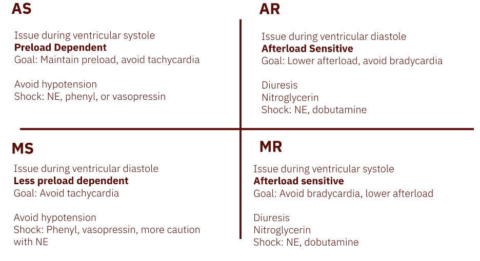
Stenotic lesions (aortic stenosis and mitral stenosis) generally require preservation of preload and adequate filling time. Hypotension should be avoided, fluids should be administered cautiously, and tachyarrhythmias should be treated promptly, as they reduce ventricular filling and cardiac output.
Regurgitant lesions (mitral and aortic regurgitation) are primarily afterload-sensitive states. The goal is to maximize forward cardiac output by reducing systemic vascular resistance whenever appropriate, avoiding unnecessary vasoconstriction, and recognizing that bradycardia often worsens regurgitation by prolonging the time available for blood to flow backward across the valve.
POCUS has become an invaluable bedside tool for identifying structural valve abnormalities and raising suspicion for acute valvular pathology. However, ultrasound findings should always be interpreted alongside the patient’s clinical presentation, as normal-appearing valves do not exclude severe disease and Doppler findings alone cannot reliably quantify severity.
Finally, remember that many of our usual reflexes in the resuscitation room can be harmful in patients with acute valvular disease. Before reaching for fluids, vasopressors, or the laryngoscope, pause to consider the underlying physiology. The most important question is often not “What is the diagnosis?” but rather, “What does this ventricle need?”
Take-home Points
- Stenotic lesions need preload, perfusion, and time to fill.
- Regurgitant lesions need forward flow and reduced afterload.
- Heart rate matters, but the optimal target depends on the valve lesion.
- POCUS should support, not replace, clinical judgment.
- When in doubt, think physiology before reflexively giving fluids, vasopressors, or proceeding with intubation.
References
- van Diepen S, Katz JN, Albert NM, Henry TD, Jacobs AK, Kapur NK, et al. Contemporary management of cardiogenic shock: a scientific statement from the American Heart Association. Circulation. 2017;136(16):e232–68.
- Thiele H, Ohman EM, de Waha-Thiele S, Zeymer U, Desch S. Management of cardiogenic shock complicating myocardial infarction: an update 2019. Eur Heart J. 2019;40(32):2671–83.
- Keane MG. Acute heart valve emergencies. Heart. 2024;110(4):247–256. (3)
- Otto CM, Nishimura RA, Bonow RO, Carabello BA, Erwin JP III, Gentile F, et al. 2020 ACC/AHA guideline for the management of patients with valvular heart disease. J Am Coll Cardiol. 2021;77(4):e25–197. (4)
- Lindman BR, Clavel MA, Mathieu P, Iung B, Lancellotti P, Otto CM, et al. Calcific aortic stenosis. Nat Rev Dis Primers. 2016;2:16006. (5)
- Otto CM, Nishimura RA, Bonow RO, Carabello BA, Erwin JP, Gentile F, et al. 2024 ACC/AHA guideline for the management of valvular heart disease. J Am Coll Cardiol. 2024;83(18):e1–e227. (6)
- Osman M, Syed M, Patibandla S, et al. Outcomes of patients with cardiogenic shock treated with mechanical circulatory support: a contemporary meta-analysis. Am J Emerg Med. 2024;72:1–9. (7)
- Vincent JL, De Backer D. Circulatory shock. N Engl J Med. 2013;369(18):1726–34. (8)
- Enriquez-Sarano M, Akins CW, Vahanian A. Mitral regurgitation. Lancet. 2009;373(9672):1382–1394. (9)
- Dziadzko V, Clavel MA, Dziadzko M, Medina-Inojosa JR, Michelena HI, Maalouf J, et al. Outcome and undertreatment of mitral regurgitation: a community cohort study. Lancet. 2018;391(10124):960–969. (10)
- Tribouilloy C, Rusinaru D, Szymanski C, Meziani F. Acute mitral regurgitation. Heart. 2021;107(14):1093–1100. (11)
- Dudzinski DM, Hung J. Echocardiographic assessment of acute valvular regurgitation. Circulation. 2014;129(25):e521–e546. doi:10.1161/CIRCULATIONAHA.112.113993 (12)
- Zoghbi WA, Adams D, Bonow RO, Enriquez-Sarano M, Foster E, Grayburn PA, et al. Recommendations for noninvasive evaluation of native valvular regurgitation. J Am Soc Echocardiogr. 2017;30(4):303–371. (13)
- Abraham WT, Adams KF, Fonarow GC, Costanzo MR, Berkowitz RL, LeJemtel TH, et al. In-hospital mortality in patients with acute decompensated heart failure requiring intravenous vasoactive medications. J Am Coll Cardiol. 2005;46(1):57–64.(14)
- Levy P, Compton S, Welch R, Delgado G, Jennett A, Penugonda N, et al. Treatment of severe decompensated heart failure with high-dose intravenous nitroglycerin: a feasibility and outcome analysis. Ann Emerg Med. 2007;50(2):144–152. (15)
- Wang K, Guo H, Zhang Z, Liu M. Role of high-dose intravenous nitrates in hypertensive acute heart failure: a systematic review. Am J Emerg Med. 2020;38(8):1662–1668.(16)
- Afilalo J, Lauck S, Kim DH, Lefèvre T, Piazza N, Lachapelle K, et al. Frailty in older adults undergoing aortic valve replacement. J Am Coll Cardiol. 2017;70(6):689–700. (17)
- January CT, Wann LS, Calkins H, Chen LY, Cigarroa JE, Cleveland JC Jr, et al. 2019 AHA/ACC/HRS focused update on atrial fibrillation management. Circulation. 2019;140(2):e125–51.(18)