
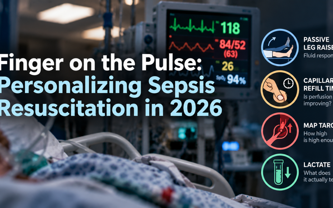
Sepsis resuscitation has evolved considerably over the past two decades, moving away from rigid protocols toward more individualized care. Yet many familiar targets remain: 30 mL/kg of fluid, a MAP of 65, and lactate clearance.
But not every patient benefits from more fluid, a higher blood pressure, or chasing a lactate to normal. In 2026, the focus is increasingly on dynamic reassessment and personalized resuscitation. This post reviews what that looks like at the bedside, and what to do after the first litre.
The Case
- Jeff: 68M, previously healthy. BP 84/52, HR 118, RR 26. WBC 24.6, Cr 187 (baseline 100), lactate 4.8 mmol/L.
- Diagnosis: Septic shock. Antibiotics started, 30 cc/kg given, vasopressors initiated, ICU called.
- The question: What do you do with him for the next hour while he’s still in your department?
Jeff anchors every concept in this talk — fluid strategy, MAP targets, lactate, and the ANDROMEDA-SHOCK 2 trial all answer the question of what happens after the first litre.
Where We Came From: The EGDT Era
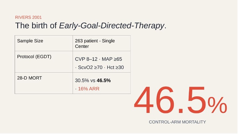
Figure 1. Rivers 2001 — The birth of EGDT. A 16% ARR in mortality, but a 46.5% control-arm rate no longer reflects modern practice.
- Rivers 2001¹: Protocolized sepsis bundle – CVP targets, ScvO2 monitoring, rigid Hgb targets showed 16% ARR in 28-day mortality vs. usual care.
- Control-arm mortality was 46.5%, far above what we see in modern practice, suggesting major baseline care improvements have already occurred.
- Three large RCTs followed (PROCESS, ARISE, ProMISe – 2014–2015)²⁻⁴: >4,000 patients across three continents. No mortality benefit over usual care. More resource-intensive monitoring required.
- What EGDT left us: A culture of chasing numbers rather than treating the patient.
- What survived: Early antibiotics, source control, and vasopressors. The specific CVP/ScvO2 targets were abandoned.
Fluids – Are We Still Living in 2001?
The 30cc/kg Dogma
- Where it came from: Expert consensus extrapolated from Rivers, not a randomized trial. Codified into order sets.
- SSC 2026 now labels this a conditional recommendation with low certainty evidence.⁵
- For an 80 kg patient: that’s still 2.4 L before any formal reassessment. Ask yourself: is this patient going to benefit from all of it?
CLASSIC and CLOVERS: Restrictive Strategies Are Safe
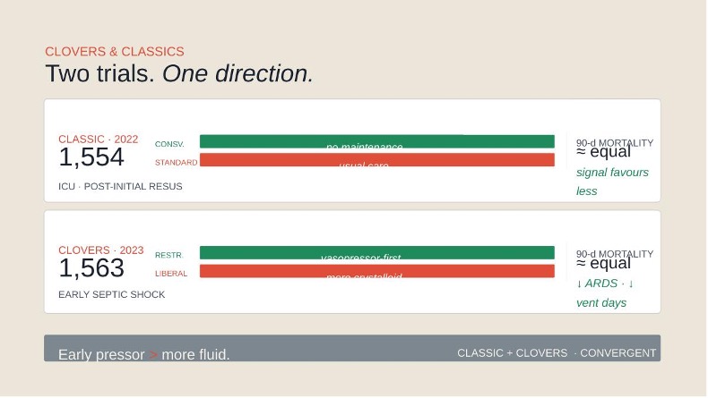
Figure 2. CLASSIC (2022) and CLOVERS (2023) — Two trials, one conclusion: restrictive fluid strategies and early vasopressors are safe.
- CLASSIC (2022)⁶: 1,554 ICU patients post-initial resuscitation. Restrictive arm: 1.8 L vs. 3.8 L in 24h. 90-day mortality: no difference.
- CLOVERS (2023)⁷: 1,563 ED patients with early septic shock. Restrictive arm used earlier vasopressors instead of more fluid. Fluid accumulation: 3.4 vs. 5.5 L. No mortality difference, with a signal toward less ARDS and fewer ventilator days.
- Key insight: Many CLOVERS control-arm patients eventually needed pressors anyway, early vasopressor initiation is not failure. It is appropriate escalation.
Why Half of Patients Don’t Respond to Fluid
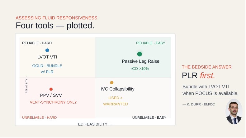
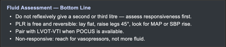
Figure 3. Assessing fluid responsiveness. Four tools plotted by reliability and ED feasibility. PLR first; bundle with LVOT-VTI when POCUS is available.
- ~50% of hemodynamically unstable patients are not fluid-responsive.⁸ Fluid in a non-responder adds harm; volume overload, ARDS, AKI – with no haemodynamic benefit.
- Three separate questions: (1) Is the patient fluid-responsive? (2) Can they tolerate more fluid? (3) Is there active hypoperfusion? Do not conflate them.
- Passive leg raise (PLR): Lay flat, raise legs 45°, watch for 10–15 mmHg SBP or 5–10 mmHg MAP rise. Free, reversible, requires no equipment.
- Gold standard: PLR + LVOT-VTI to quantify cardiac output response.
- IVC collapsibility: A plethoric IVC reliably rules OUT fluid responsiveness. A collapsing IVC does not rule it in. Do not use as a sole decision-maker.
- PPV: Requires mechanical ventilation and sedation, limited utility in the ED.
“PLR is fairly robust and reliable, it’s not the easiest thing to do, but it tells you something real. Pick a threshold that’s meaningful for the person in front of you and pair it with what their skin is telling you.” – Dr. Rakesh Patel, Intensivist, University of Ottawa
MAP Targets – Is 65 Sacred?
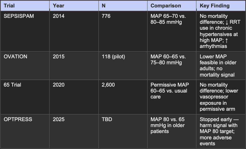
Table 1. MAP target RCTs – four trials, one direction. Higher is not better.
- OPTPRESS (2025)¹⁰: Stopped early – increased adverse events in the MAP 80 arm, even in older hypertensive patients previously thought to benefit from higher perfusion pressure.
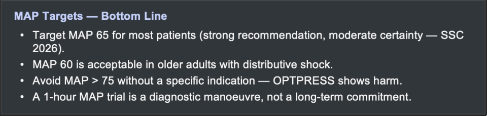
- SSC 2026: Strong recommendation with moderate certainty for MAP 65. MAP 60 explicitly accepted in older adults with distributive shock.⁵
- Clinical application: A time-limited 1-hour trial at MAP 60 is evidence-supported for borderline patients before committing to vasopressor escalation.
“The 2026 guidelines explicitly accept MAP 60 in older patients with distributive shock. OPTPRESS reinforces that aggressive MAP targets carry real risk, more vasopressor exposure is not neutral.” – Dr. Bram Rochwerg, SSC 2026 Vice-Chair, McMaster University
Lactate – The Most Misunderstood Lab in Sepsis
- Three mechanisms elevate lactate: (1) Anaerobic production from tissue hypoperfusion, (2) catecholamine-driven aerobic lactate production (endogenous stress hormones or exogenous beta-2 agonists), (3) decreased hepatic clearance. These coexist in every septic patient.
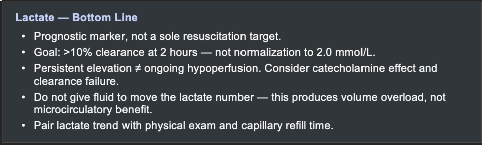
- Attributing an elevated lactate solely to hypoperfusion and responding with more fluid is a common and potentially harmful error.
Clearance, Not Normalization
- Goal: >10% clearance at 2 hours¹¹ – independently associated with improved survival.
- SSC 2026: Serial lactates should target decrement, not normalization. Continuing fluid until lactate reaches 2.0 mmol/L leads to volume overload without microcirculatory benefit.⁵
- Lactate clearance can fall due to dilution just as easily as because perfusion has improved; the dynamic is hard to interpret in isolation.
- Best paired with: Physical exam, capillary refill time, and clinical trajectory.
“A high lactate is not a prescription for more fluid. It’s a signal that something is wrong — and your job is to figure out what.” – Dr. Rakesh Patel, Intensivist, University of Ottawa
The Microcirculation – Why the Macro Is Not Enough
- Macro-only monitoring fails: You can normalize MAP, clear lactate, and still have ongoing microcirculatory dysfunction driving end-organ failure.
- Septic shock is not simply low blood pressure; it is maldistribution of flow at the microvascular level: endothelial dysfunction, microthrombus formation, and loss of autoregulation.¹²
- CRT is the practical bedside tool for tracking microcirculatory response to resuscitation.
CRT – Standardized Technique
- Location: Ventral surface of the right index finger.
- Apply 10 seconds of continuous, firm pressure.
- Time the return of colour with a stopwatch or phone timer.
- Target: Normalization within 3 seconds.
- Use a timer. Clinicians systematically undercount without one. A phone stopwatch takes 2 seconds and eliminates the most common measurement error.
ANDROMEDA-SHOCK 2 – CRT Meets the RCT
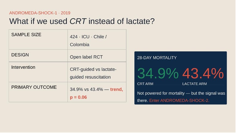
Figure 4. ANDROMEDA-SHOCK 1 — CRT- vs. lactate-guided resuscitation. Mortality trended lower (34.9% vs. 43.4%, p=0.06) but was underpowered. Generated the hypothesis for AS-2.
- Design: 424 patients, open-label RCT, Chile/Colombia. Septic shock randomized to CRT-guided vs. lactate-guided escalation.¹³
- Result: 28-day mortality 34.9% (CRT) vs. 43.4% (lactate), p=0.06 – trend, not significant. Underpowered for mortality.
- Conclusion: Generated a clear hypothesis: targeting the microcirculation directly may be superior to using lactate as a surrogate. → ANDROMEDA-SHOCK 2.
ANDROMEDA-SHOCK 2 — Study Design
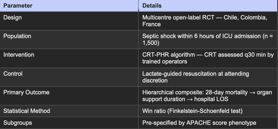
Table 2. ANDROMEDA-SHOCK 2 study design. Hernandez et al., NEJM, 2025.
Understanding the Win Ratio
- Each intervention patient is paired head-to-head with each control patient.
- Hierarchy: (1) Did one patient die at 28 days? → they lose. (2) If both survived, who had shorter organ support duration? (3) Still tied → shorter hospital stay wins.
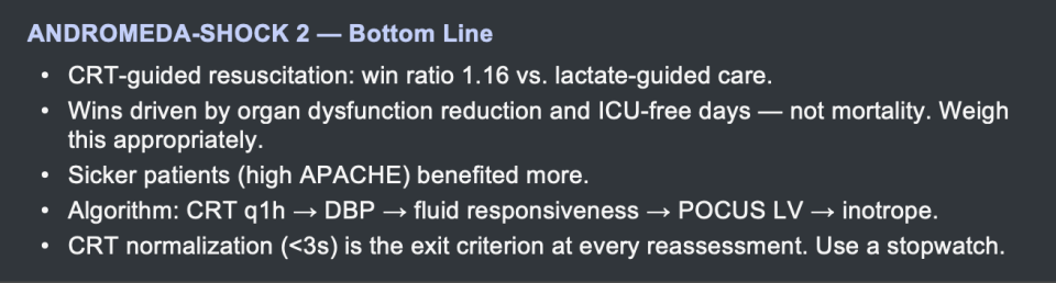
- Win ratio = 1.16 — CRT-guided patients won 54 times per 100 pairs vs. 46 losses. In high-APACHE patients, benefit was more pronounced.
- Isolated 28-day mortality: HR 0.99, p = 0.91 – no difference. Wins were driven by reduced organ dysfunction and more ICU-free days.
- Interpretation: Whether organ dysfunction endpoints alone justify practice change is debatable. A mortality signal from squeezing a finger was a tall ask, the organ burden data are still meaningful.
The Simplified CRT-PHR Algorithm for the ED
After initial resuscitation (fluids, vasopressors, antibiotics), reassess at minimum every hour using this framework:
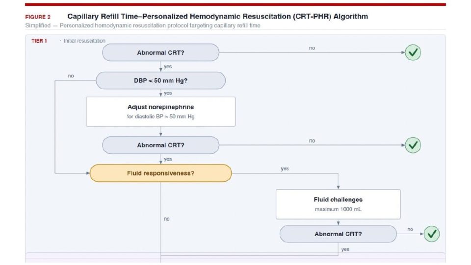
Figure 5. CRT-PHR Algorithm — Tier 1. Start with CRT. Normalized (<3s): reassess in 1 hour. Abnormal: check DBP first, then fluid responsiveness. (Simplified from Hernandez et al., 2025) https://jamanetwork.com/journals/jama/fullarticle/2840823
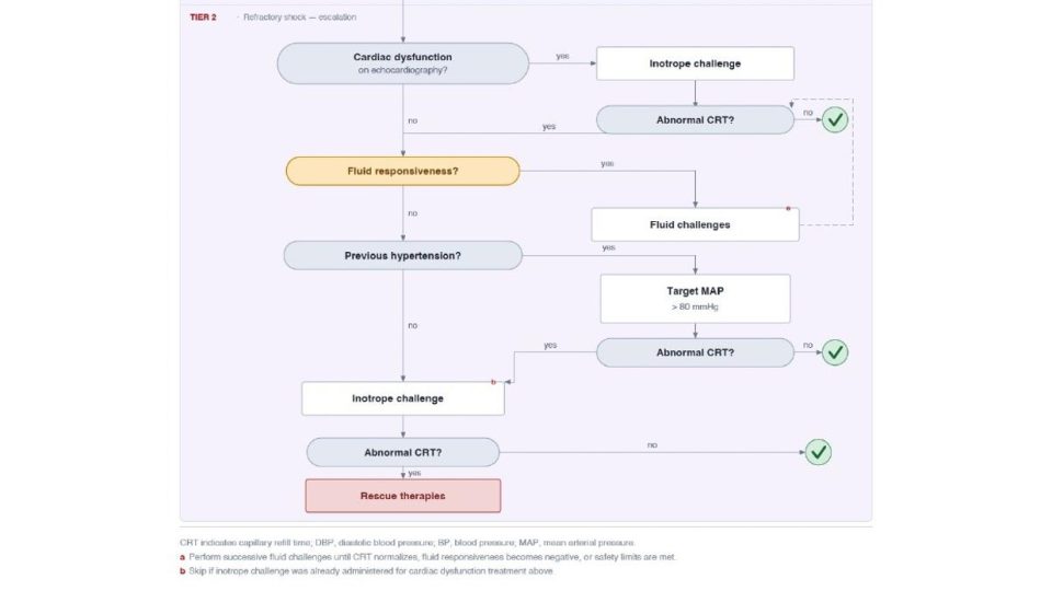
Figure 6. CRT-PHR Algorithm — Tier 2 (Refractory Shock). POCUS LV assessment → inotrope if LV down → MAP trial at 80 mmHg if prior hypertension → rescue therapies if CRT remains elevated. https://jamanetwork.com/journals/jama/fullarticle/2840823
Tier 1 – every reassessment:
- CRT ≥ 3s? → Check diastolic BP first. DBP < 50 → adjust norepinephrine or add vasopressin to target DBP > 50.
- DBP adequate + CRT still abnormal? → Assess fluid responsiveness (PLR, VTI, or 250 mL test bolus).
- Fluid-responsive: Give challenges up to 1 L while rechecking CRT.
- Non-responsive: → Enter Tier 2.
Tier 2 – refractory shock:
- POCUS LV assessment (qualitative eyeball — is LV up or down?).
- LV dysfunction: → Add dobutamine 5 mcg/kg/min (or low-dose epinephrine as accessible alternative).
- No LV dysfunction + prior hypertension: → 1-hour MAP trial at 80 mmHg, recheck CRT.
- CRT still abnormal after both: → ICU escalation; you have exhausted your ED toolkit.
“Personalized resuscitation for septic shock is a superior strategy to protocol-driven care. It’s important to incorporate dynamic, patient-specific indices when managing critically ill patients — even in a busy ED.” – Dr. Kevin Durr, EM/CC Physician, University of Ottawa
Jeff – What This Looked Like
- Hour 1: CRT 4s. DBP 42. → Increase norepinephrine, add vasopressin. Recheck.
- Hour 2: MAP 65, DBP normalized. CRT 3.5s. PLR negative. → POCUS: severely reduced LV function. → Add dobutamine 5 mcg/kg/min.
- Hour 3: BP 100/54 (MAP 69). CRT 2.5s. Lactate 2.7 and trending down. The patient now has a plan.
Surviving Sepsis Campaign 2026 – What Changed
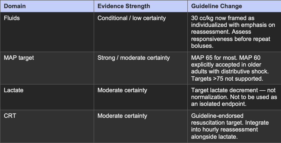
Table 3. SSC 2026 — Key changes for emergency medicine practice.
- The biggest conceptual shift: From “how much fluid did we give?” to “did the patient actually respond?” Dynamic reassessment tools are now explicitly endorsed over fixed-volume protocols.
Three Questions at Every Reassessment
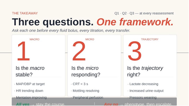
Figure 7. Three Questions. One Framework. Ask at every bedside reassessment — minimum every hour.
- Q1 – Is the macro stable? MAP/DBP at target. HR trending down. Mentation improving.
- Q2 – Is the micro responding? CRT < 3 seconds. Mottling resolving. Peripheries warming.
- Q3 – Is the trajectory right? Lactate decreasing. Urine output improving. Pressors weaning.
- All three yes: Stay the course.
- Any one no: Phenotype, investigate, escalate appropriately.
Your finger and your phone are already in the room. The only thing missing is the habit.
Six Things to Take to Your Next Shift
- Fluid is a drug. Assess responsiveness with PLR before every repeat bolus.
- MAP 65 is the target, but MAP 60 is safe in older adults with distributive shock. Don’t chase higher without evidence.
- High lactate is a signal, not a fluid prescription. Track trend; target >10% clearance at 2 hours.
- Check CRT with a stopwatch. Target < 3s. Dorsal right index finger, 10 seconds of firm pressure.
- The AS-2 algorithm is feasible in the ED: CRT → DBP → fluid responsiveness → POCUS LV → inotrope.
- Your septic shock patients are the sickest in the department. They deserve a bedside reassessment every hour — not just a phone call to ICU.
About the Author
Naman Arora is a PGY-4 Emergency Medicine resident at the University of Ottawa at the time of this post. He presented this Grand Rounds on May 7, 2026, under the supervision of Dr. Ariel Hendin. He would like to thank Dr. Bram Rochwerg, Dr. Rakesh Patel, and Dr. Kevin Durr for their expert contributions to this talk, as well as his partner Nikki for her work on the presentation design.
References
- Rivers E, et al. Early goal-directed therapy in the treatment of severe sepsis and septic shock. N Engl J Med. 2001;345(19):1368–1377.
- Yealy DM, et al. (ProCESS) A randomized trial of protocol-based care for early septic shock. N Engl J Med. 2014;370(18):1683–1693.
- ARISE Investigators. Goal-directed resuscitation for patients with early septic shock. N Engl J Med. 2014;371(16):1496–1506.
- Mouncey PR, et al. (ProMISe) Trial of early, goal-directed resuscitation for septic shock. N Engl J Med. 2015;372(14):1301–1311.
- Evans L, et al. Surviving Sepsis Campaign: International Guidelines 2026. Intensive Care Med / Crit Care Med. 2026.
- Meyhoff TS, et al. (CLASSIC) Restriction of intravenous fluid in ICU patients with septic shock. N Engl J Med. 2022;386(26):2459–2470.
- Semler MW, et al. (CLOVERS) Liberal versus restrictive intravenous fluid therapy in early septic shock. N Engl J Med. 2023;388(10):881–891.
- Cecconi M, et al. Fluid challenges in intensive care: the FENICE study. Intensive Care Med. 2015;41(9):1529–1537.
- Muller L, et al. Respiratory variations of inferior vena cava diameter to predict fluid responsiveness. Crit Care. 2012;16(5):R188.
- Jonsson AB, et al. (OPTPRESS) Higher versus lower MAP targets in patients aged ≥65 with septic shock. Intensive Care Med. 2025.
- Nguyen HB, et al. Early lactate clearance is associated with improved outcome in severe sepsis and septic shock. Crit Care Med. 2004;32(8):1637–1642.
- De Backer D, et al. Microcirculatory alterations in patients with severe sepsis. Crit Care Med. 2013;41(1):67–76.
- Hernandez G, et al. (ANDROMEDA-SHOCK-1) Effect of a resuscitation strategy targeting peripheral perfusion status vs serum lactate levels on 28-day mortality. JAMA. 2019;321(7):654–664.
- Hernandez G, et al. (ANDROMEDA-SHOCK-2) Effect of a peripheral perfusion-guided resuscitation strategy on outcomes in patients with septic shock. N Engl J Med. 2025.
- Asfar P, et al. (SEPSISPAM) High versus low blood-pressure target in patients with septic shock. N Engl J Med. 2014;370(17):1583–1593.


Excellent Review