
It is three in the morning. A 58-year-old man with hypertension walks in with sudden, severe chest pain. He looks uncomfortable but stable. His ECG is normal. His troponin is pending. And somewhere in the back of your mind, a quiet voice says: could this be a dissection?
What happens next depends almost entirely on who is working that night. And that, more than anything else, is why this talk exists.
This is not a review of the pathophysiology, the anatomy, or the classification systems of dissection, as that was previously covered here. What follows is something different. The goal is to challenge how we think about this diagnosis, to introduce a Canadian guideline that changes how we work these patients up, and to make one case above all: that what we are doing right now is not just excessive. It might be causing harm to our patients.
Why Are We So Scared?
Why does that voice show up? Why does dissection grip us the way it does? There are three reasons, and all three are completely fair.
First, the mortality is real. You have all heard the number: one to two percent per hour. But it is worth knowing where that number comes from. It comes from data from the 1950s, before modern surgery. Newer evidence puts untreated Type A mortality closer to 0.5% per hour in the first 24 to 48 hours. Still high. Still urgent. But the figure most of us carry in our heads is from a different era of medicine.
Second, the horror stories are real. Most of us have heard one or seen one. It stays with you in a way that a PE or a pneumonia never does. It becomes a reference point that is almost impossible to shake.
Third, the legal fear is real. None of us wants to be the doctor who missed this diagnosis. Missing it feels like professional failure and potential liability. That fear sits behind every decision we make. All of this is real. The question is whether the fear is calibrated. Does the size of the fear actually match the size of the risk? It does not. And that mismatch is quietly costing our patients.
Availability Bias
There is a concept in psychology called availability bias, and it is a simple idea: we judge how likely something is by how easily an example comes to mind. The more vivid the example, the more emotional it is, the more likely it feels, even when the numbers say something completely different.
Think about your own patients for a moment. They are terrified of shark attacks and plane crashes. Those things are scary. They are easy to picture. But in reality, they are far more likely to die of diabetes. The fear has nothing to do with the actual risk and everything to do with the emotion captured in those events.
We do exactly the same thing with aortic dissection. We have seen the case, heard the story, or sat through an M&M round where someone missed it. That image burns itself in, and from that moment on, every chest pain patient carries a shadow of that case. We order the CT, not because the pre-test probability calls for it, but because the weight of that worst case is too heavy to put down.
Dramatic morbidity. High stakes. Unforgettable cases. But prevalence is roughly 0.1%. That is availability bias. And naming it, really naming it, is the first step to correcting it.
Dissection in Context: What the Numbers Actually Say
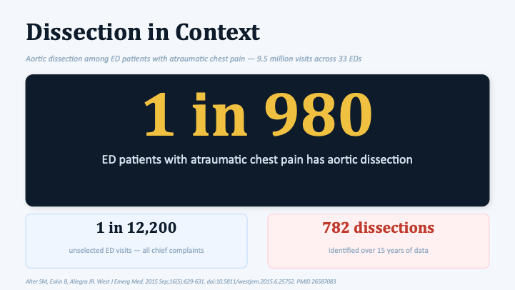
Alter and colleagues looked at 9.5 million emergency department visits across 33 hospitals. Over 15 years, they identified 782 aortic dissections. That works out to:
- 1 in 980 ED patients with atraumatic chest pain has an aortic dissection.
- 1 in 12,200 unselected ED visits, across all chief complaints.
One in nine hundred and eighty. That is your real pre-test probability. Not one in ten. Not one in a hundred.
Here is how to use that number. Because aortic dissection is a low-prevalence disease, your starting point should be low. You begin with a low baseline estimate and then adjust, up or down, based on the clinical features your patient actually has. For most of us, our gut feeling does not match the data. That difference is availability bias.
What Actually Walks Through Your Door
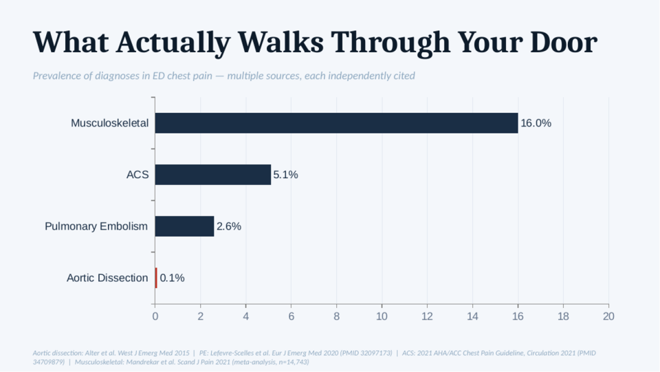
Each bar on this chart comes from a separate study, but together they tell a consistent story about what chest pain really looks like when it walks through your door.
- Musculoskeletal: 16%. About one in six chest pain patients, from a systematic review and meta-analysis of nearly 15,000 patients.
- Acute coronary syndrome: 5.1%. The diagnosis we are most trained to find, from the 2021 AHA/ACC chest pain guideline.
- Pulmonary embolism: 2.6%. And that is only among patients who actually had a workup, so the true rate is probably even lower.
- Aortic dissection: 0.1%. One tenth of one percent. That bar is barely visible on the chart. And that is the diagnosis driving the fear in the room.
Look at that chart honestly. Does your practice look like it is calibrated to one tenth of one percent? If we are being honest, most of us practice as if the number were closer to ten.
Even the CMPA Expects You to Miss It
The numbers are interesting, but what about the legal risk? What if I am the one who misses this diagnosis?
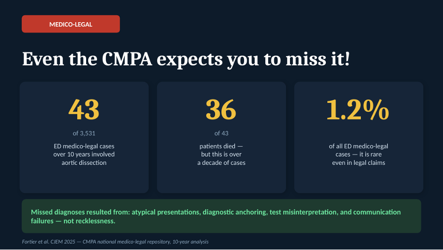
Fortier and colleagues published a ten-year analysis of the entire national CMPA medico-legal database in CJEM in 2025, the largest dataset of its kind in this country. Out of 3,531 emergency department medico-legal cases over a full decade, just 43 involved aortic dissection. That is 1.2% of all ED medico-legal cases for the one condition we often feel compelled to scan everyone for.
It is true that 36 of those 43 patients died. But hold that number carefully. Thirty-six deaths over ten years, across the whole country, tells you how severe this disease is when it is missed. It does not tell you how often missing it leads to legal trouble. Those are two very different things.
And when the authors asked why these cases were missed, the answer was not carelessness or negligence. It was atypical presentations, diagnostic anchoring, test misinterpretation, and communication failures. These are understandable errors in a difficult disease, not recklessness.
This is not permission to be sloppy. It is permission to stop practicing in fear.
It is worth sitting with why this fear is so powerful because the incentives are not balanced. The harms of over-testing are diffuse and quiet: radiation, incidental findings, cost, longer ED stays, and pressure on beds. None of that lands on the physician who ordered the scan. None of it is visible. The harm of a missed diagnosis is the opposite: vivid, personal, and sometimes accompanied by a lawsuit.
So, it is entirely rational that we lower our threshold to test, even when the benefit is tiny. But that instinct does not always line up with good, patient-centred care. When we treat the patient in front of us and their actual presentation, we are giving high-quality care. When we treat our own anxiety, we start giving worse care, care that is no longer centred on the patient.
Acute Aortic Syndrome in Our Own Backyard
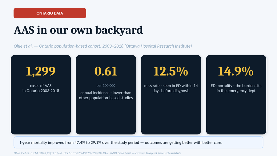
Ohle and colleagues studied every case of acute aortic syndrome in Ontario between 2003 and 2018, representing fifteen years of provincial data, co-authored here at the Ottawa Hospital Research Institute.
- 1,299 cases of AAS in the entire province across 15 years.
- 0.61 per 100,000 annual incidence, lower than in other population-based studies. Even in Ontario, this condition is less common than many of us expect.
- 12.5% miss rate, defined as patients seen in an ED within 14 days before the diagnosis. That is about one in eight, not most patients.
- 14.9% ED mortality. The burden of death sits in the emergency department. That is exactly why a talk like this matters, because that is where we work.
One-year mortality also improved over the study period, from 47.4% to 29.1%. The authors are careful not to claim a single cause, and neither will I. Better surgery, earlier recognition, or a different case mix may have contributed; we cannot say for certain. What we can say is that outcomes are getting better with better care.
So, the picture from Ontario is fairly clear: dissection is uncommon, missed cases do occur, and outcomes have improved substantially over time. The question we have to ask ourselves is whether we are practising according to the evidence or according to our fears.
And in Our Own Department?
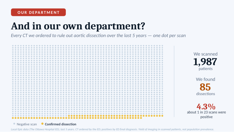
Every CT ordered to rule out aortic dissection at The Ottawa Hospital ED over the last five years, with one dot per scan. Local Epic data; CTs ordered by the ED, positives by ED final diagnosis. This is the yield of imaging in scanned patients, not population prevalence.
Everything above comes from the published evidence: national numbers, provincial numbers. But before going any further, it is worth asking a simple question: what does this look like in our own department at The Ottawa Hospital?
We reviewed every CT ordered to rule out aortic dissection over the past five years. Each dot on that image is one scan.
- 1,987 patients scanned.
- 85 dissections found.
- 4.3% positive rate, about 1 in 23 scans. Which means more than 95% of these scans were negative.
To be clear about what this number means: it reflects the yield among patients we had already decided to scan. It is not the prevalence among all patients presenting with chest pain. But that is exactly the point. When we cast the net this wide, most of what we catch will be negative: a large wall of grey and only a thin band of gold.
So, if most of what we are catching is grey, what does that actually cost the patient? Take radiation as an example. A CT of the aorta delivers roughly 15 to 20 millisieverts. In practical terms, that is about five to seven years of natural background radiation delivered in a few seconds. Over the last five years, we have delivered that dose almost two thousand times to find 85 dissections.
This is not a judgment of anyone. I have ordered plenty of these scans myself. But it is worth stepping back and looking at the whole picture. Every one of those grey dots came with a cost: contrast, radiation, incidental findings, time in the department, and often downstream testing as well.
What strikes me is how differently we talk about this risk. When a patient asks for a scan we do not think they need, we are often very careful to explain the radiation. But when we are the ones ordering the scan, that same risk somehow fades into the background.
Your Colleagues Have Already Decided
If all of that is still not enough, Ohle and colleagues went straight to your peers. They ran a national survey of Canadian emergency physicians asking one simple question: what miss rate for aortic dissection would you personally find acceptable?
Two in three Canadian emergency physicians accept a miss rate of less than 1%. Not zero. Less than one percent. The profession itself has already made peace with this reality: dissection is rare, it presents in many different ways, and a zero-miss rate is simply not achievable. The question is whether our day-to-day practice reflects that.
So, What Is the Fear Actually Doing to Our Practice?
If fear is driving the conversation, the next question is simple: what is it doing at the bedside?
- Over-investigation: exposing people to radiation they did not need.
- Incidental findings: lung nodules that generate years of follow-up scans and years of worry for something that will never cause harm.
- Contrast nephropathy: contrast injury to the kidneys.
- Downstream harm: testing cascades that nobody intended and nobody benefits from.
The key message is this: testing is not a neutral act. Every time we order a test, we make a decision that exposes patients to risks and downstream consequences, even when the result is negative.
There is also a subtler cost. The more we scan by reflex, the less we use our own clinical judgment. We drift from probability-based medicine toward possibility-based medicine, from asking, “How likely is this?” to asking, “Could this be possible?” And almost anything is possible.
The harms of over-testing belong largely to the patient and the healthcare system. The reassurance belongs largely to the clinician.
Some readers will be thinking: if one in eight dissections were missed in the Ontario study, should we not be scanning more? It is a fair question, but I do not think so. The goal is not to miss fewer dissections by scanning indiscriminately; the goal is to find the right dissections. The missed cases are often atypical presentations that never entered the diagnostic pathway in the first place. Scanning more low-risk chest pain patients will not find them. Better clinical reasoning will. More scans do not automatically mean more accuracy.
What Actually Moves the Needle?
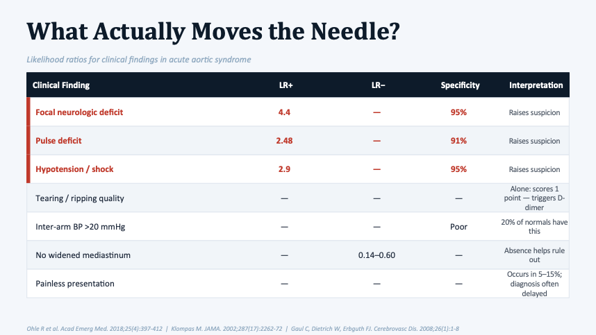
The message here is simple: many of the classic findings we associate with aortic dissection do not perform the way we think they do.
- Focal neurologic deficit: LR+ 4.4, specificity 95%. Raises suspicion.
- Pulse deficit: LR+ 2.48, specificity 91%. Raises suspicion.
- Hypotension or shock: LR+ 2.9, specificity 95%. Not common, but when present, it should raise your suspicion.
According to the Ohle review, those are the three findings that truly move the needle.
Tearing or ripping quality. Despite its reputation, the evidence is mixed. The studies were too inconsistent for the Ohle review to draw a firm conclusion. That does not make it useless. Tearing pain remains a high-risk pain feature in the Canadian guideline, so a patient with tearing pain scores a point on the Canadian tool and qualifies for D-dimer testing. It belongs in your assessment; it just cannot carry the diagnosis by itself.
Inter-arm BP difference >20 mmHg. Poor specificity: 20% of people without dissection have this. More on that below.
No widened mediastinum: LR− 0.14–0.60. This is the one finding that points in the opposite direction. A normal mediastinum on chest X-ray does not rule out dissection, but it does make it less likely and can be useful as part of a low-risk presentation.
Painless presentation. Occurs in 5–15%, and diagnosis is often delayed. These are the patients who arrive with stroke symptoms, syncope, abdominal pain, or limb ischemia, and they are the cases most likely to be missed.
On those painless presentations, Dr. Shahbaz Syed (ED Physician at TOH) makes an important point: if a patient has a dissection but no pain and no features that suggest the diagnosis, some cases will inevitably be missed. We cannot be expected to diagnose a disease when there is nothing in the presentation pointing us toward it. The goal is not perfection against the impossible. The goal is to recognize the patients who give us something to work with.
Bilateral Blood Pressure Differential: Stop Doing This
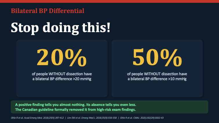
When we worry about dissection, we are taught to check the blood pressure in both arms. That habit deserves a challenge because the evidence suggests it is not as helpful as many of us think.
- 20% of people WITHOUT dissection have a bilateral BP difference greater than 20 mmHg.
- 50% of people WITHOUT dissection have a difference greater than 10 mmHg.
So, what happens when you find a difference? Not much. It does not meaningfully make dissection more likely. And what if you do not find one? That does not help either; a normal reading in both arms does not make dissection less likely. A positive finding tells you almost nothing, and its absence tells you even less.
This is why the Canadian guideline removed blood pressure differential from its list of high-risk features, and it is the only major guideline to do so. The reasoning is exactly what the evidence above shows.
A study by Um, Ohle, and Perry in 2018 looked at this sign specifically in the emergency department. Their conclusion was clear: the difference between arms is statistically linked to dissection, but it adds nothing once you have already checked the pulse properly. It does not improve your accuracy. It is time we stopped giving this finding more weight than it deserves.
The ADD Score
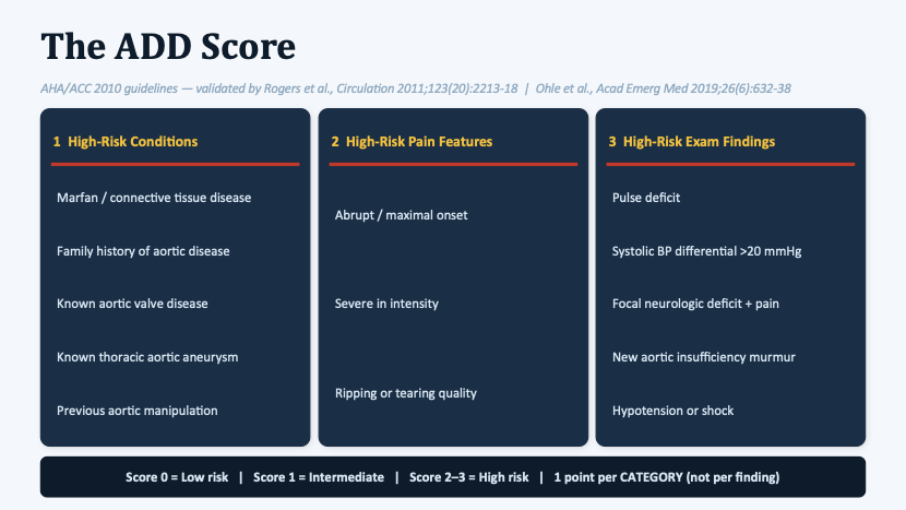
The ADD-RS, or Aortic Dissection Detection Risk Score, is the tool most of us use to assess how likely a dissection is. It was introduced in the 2010 thoracic aortic guidelines and validated by Rogers and colleagues in 2011. It has three categories:
- High-risk conditions: Marfan or connective tissue disease, family history of aortic disease, known aortic valve disease, known thoracic aortic aneurysm, previous aortic manipulation.
- High-risk pain features: abrupt or maximal onset, severe in intensity, ripping or tearing quality.
- High-risk exam findings: pulse deficit, systolic BP differential >20 mmHg, focal neurologic deficit with pain, new aortic insufficiency murmur, hypotension or shock.
A score of 0 is low risk, 1 is intermediate risk, and 2–3 is high risk. Here is the part many people forget: you score one point per CATEGORY, not one point per finding. A patient with Marfan syndrome, tearing pain, and a pulse deficit scores three, not five.
The score has one key limitation. A score of 1 or more identifies about 95% of dissections, but its specificity is only about 26%. In practical terms, it flags a lot of patients. So, while the score is good at identifying who might have a dissection, it does not tell you who needs a CT. That is exactly what D-dimer adds.
The Rule-Out Strategy: ADD-RS + D-Dimer
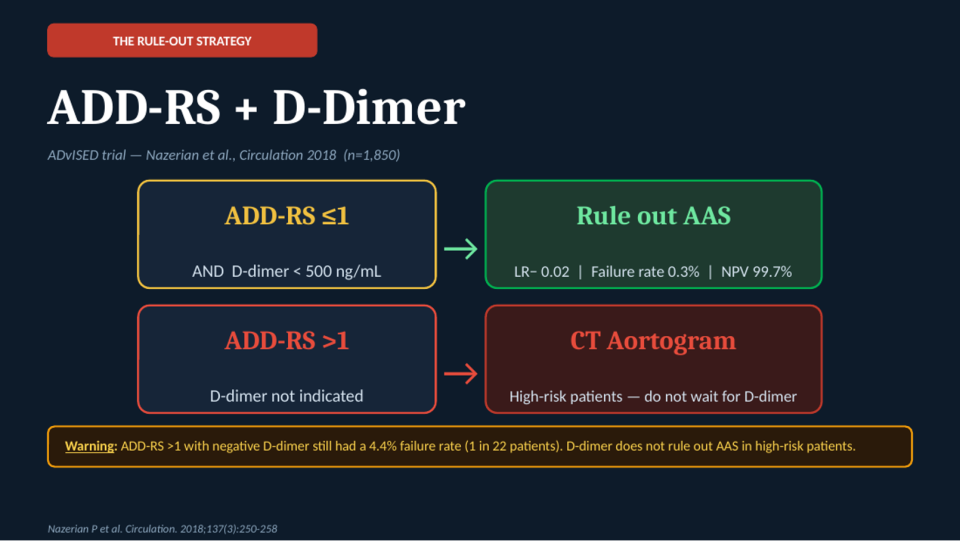
This is where it comes together. The ADvISED trial, published in 2018, was a prospective study across six hospitals in four countries, enrolling 1,850 patients in whom the treating physician was already considering acute aortic syndrome. To date, it remains the only prospective study of this combined approach.
ADD-RS ≤1 AND D-dimer <500 ng/mL → rule out AAS. LR− 0.02, failure rate 0.3%, NPV 99.7%, sensitivity 98.8%. In practical terms, roughly one missed case for every 300 patients. Even the worst end of the confidence interval was only about 1%, well below the 3% benchmark we accept for ruling out PE.
ADD-RS >1 → CT aortogram. D-dimer is not indicated. In high-risk patients, a negative D-dimer still missed five cases, a 4.4% failure rate, or about one missed case in every 22 patients. A negative D-dimer cannot safely rule out dissection in a high-risk patient.
The practical message is simple. If your patient has a pulse deficit, a neurologic deficit, hypotension, or otherwise looks high risk, skip the D-dimer. Go straight to CT.
How Does It Perform?
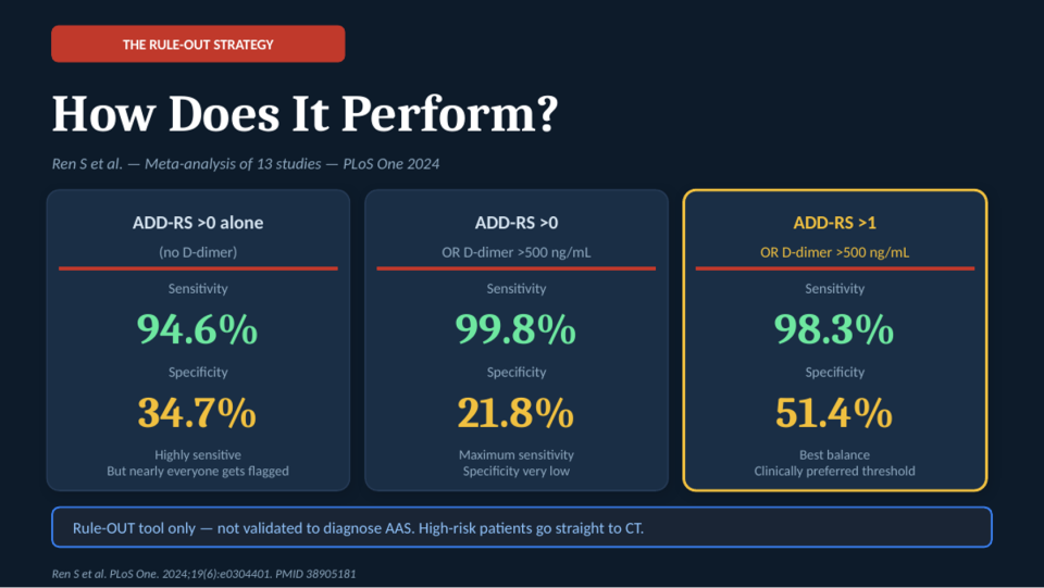
A 2024 meta-analysis by Ren and colleagues combined data from thirteen studies looking at the ADD-RS alone and in combination with D-dimer. Before walking through it, here is the key idea: you scan if either test is positive; you only rule out when both are negative.
- ADD-RS >0 alone (no D-dimer): sensitivity 94.6%, specificity 34.7%. Highly sensitive, but nearly everyone gets flagged.
- ADD-RS >0 OR D-dimer >500 ng/mL: sensitivity 99.8%, specificity 21.8%. Maximum sensitivity, but specificity is very low. You end up scanning even more patients, most of whom do not have the disease.
- ADD-RS >1 OR D-dimer >500 ng/mL: sensitivity 98.3%, specificity 51.4%. The best balance and the clinically preferred threshold. You rule out only if the score is 1 or less AND the D-dimer is negative.
One final reminder: this is a rule-OUT tool only. It has not been validated to diagnose AAS. A negative result tells you it is reasonable not to scan; it does not prove the disease is absent, and it is not intended for high-risk patients. Those patients go straight to CT.
Thinking in Probabilities
We have spent a lot of time on clinical findings, risk scores, and D-dimer. But one idea ties all of it together.
The issue is not knowledge. Everyone knows what sensitivity and specificity mean. The problem is that sensitivity and specificity only describe how a test behaves. They do not tell you what a result means for the patient in front of you. To understand that, you need one more piece of information: how likely the disease was before the test was ordered. That is pre-test probability, and it is the most important concept in this entire talk. The same test result can mean very different things in two different patients, depending on how likely the disease was to begin with.
How a Test Actually Updates You
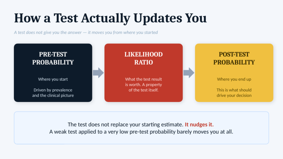
A test does not give you the answer. It does one thing: it moves your suspicion up or down.
- Pre-test probability: where you start, driven by prevalence and the clinical picture. For dissection in a typical chest pain patient, we already know that number: about one in a thousand.
- Likelihood ratio: what the test result is worth. A property of the test itself. Think of it as a multiplier for your suspicion. An LR of 5 means the finding is five times more likely in someone with the disease than without it. An LR close to 1 barely moves the needle.
- Post-test probability: where you end up. This is what should drive your decision.
The key point is that the likelihood ratio belongs to the test, not the patient. A pulse deficit has the same effect whether the patient is high risk or low risk. What changes is where you started.
The test does not replace your starting estimate. It nudges it. A weak test applied to a very low pre-test probability barely moves you at all. If you start at one in a thousand, even a positive test may still leave you with a very small probability. And if you start high, a reassuring test may not be enough to bring you down to safety.
(Strictly, the likelihood ratio multiplies the odds, not the raw probability. At the very low probabilities we are dealing with here, the two are almost the same, so “multiply your suspicion” is a fair way to picture it. The figures below are worked out properly using odds.)
What Does a Pulse Deficit Actually Do?
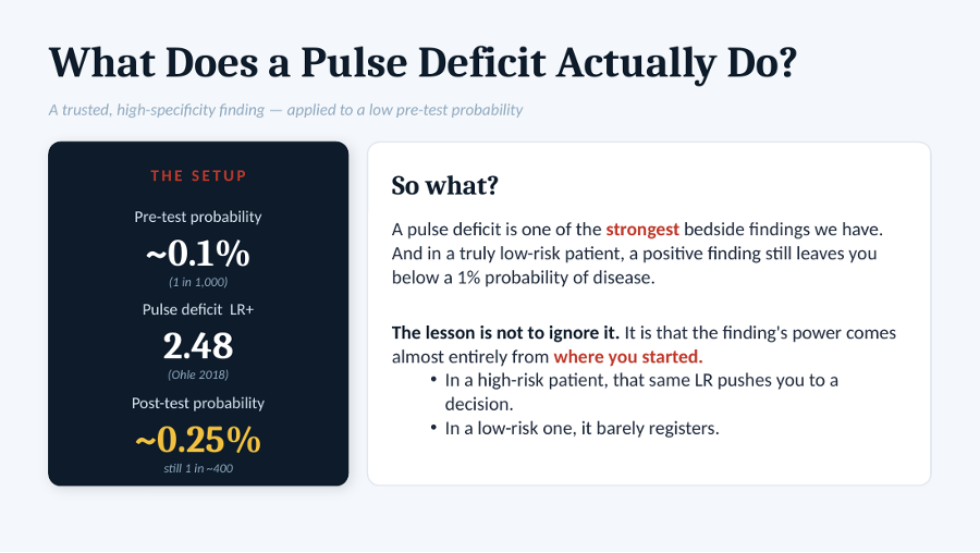
Remember the pulse deficit, one of the strongest bedside findings we have. Start with a low-risk patient whose chance of dissection is about 0.1%, or one in a thousand. Now add a pulse deficit with a likelihood ratio of about 2.48. Their probability rises to roughly 0.25%, still about one in four hundred.
That is it. Still low. Still nowhere near a diagnosis.
The lesson is not to ignore the finding. It is that the finding’s power comes almost entirely from where you started. In a high-risk patient, that same LR pushes you to a decision. In a low-risk one, it barely registers. In real life, these patients often have severe pain, a concerning story, or other high-risk features. Their starting probability was already high. The likelihood ratio is fixed. What changes is where you started.
What Does a Normal D-Dimer Buy You?
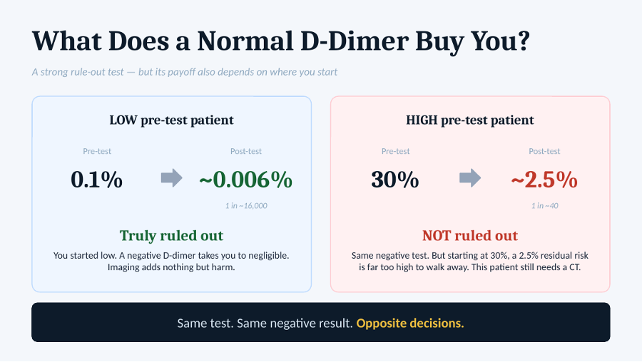
D-dimer is genuinely good at one thing: ruling out dissection. But even a very good rule-out test can only do so much.
- The low pre-test patient: starts at 0.1%. A negative D-dimer drops them to about 0.006%, roughly 1 in 16,000. Truly ruled out. Imaging adds nothing but harm.
- The high pre-test patient: imagine someone with Marfan syndrome, tearing pain, and a pulse deficit, with a starting probability around 30%. The same negative D-dimer only brings them to about 2.5%, or 1 in 40. NOT ruled out. That patient still needs a CT.
Two and a half percent is far too high to walk away from for a disease that can kill within the hour. No one is walking away from that patient because the D-dimer happened to be negative.
Same test. Same result. Completely different decisions. The only thing that changed was where the patient started, and that is exactly why the guideline does not allow a negative D-dimer to rule out dissection in a high-risk patient.
The Same Test in Two Worlds
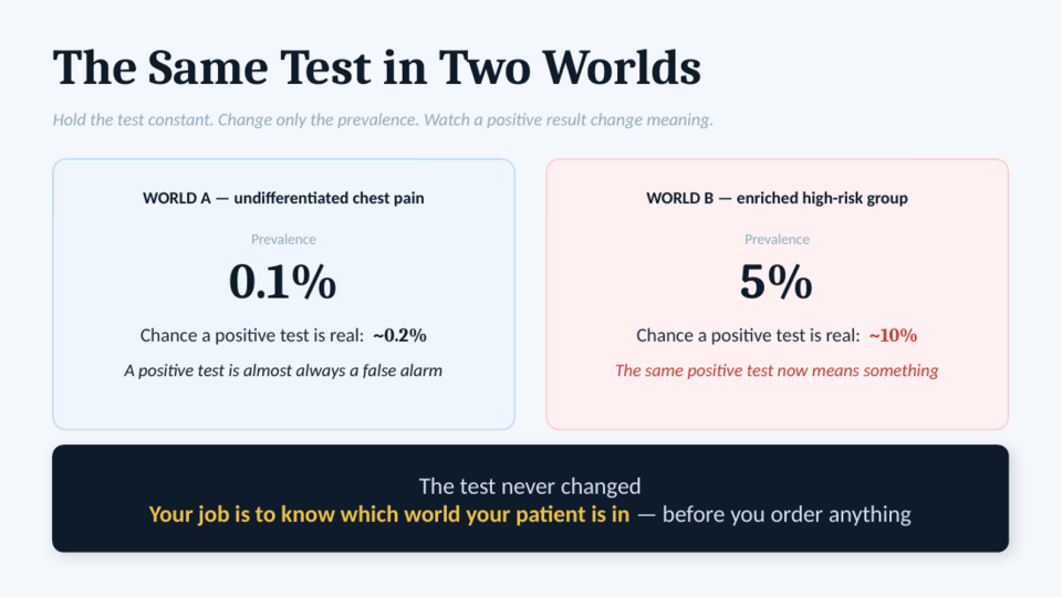
This captures the whole argument in one picture. Run the exact same test in two different worlds. Same sensitivity. Same specificity. The only thing that changes is how likely the disease is before the test.
- World A: undifferentiated chest pain, prevalence 0.1%. The chance that a positive test is real is about 0.2%. A positive test is almost always a false alarm. It triggers more testing, more scans, and more worry, but very few positives represent a true dissection.
- World B: enriched high-risk group, prevalence 5%. The chance that a positive test is real is about 10%. The same positive test now means something.
The test never changed. The patient did. A test result only makes sense in the context of the patient you are managing. Your job is to know which world your patient is in before you order anything.
Worth noting: the authors place D-dimer’s useful range at a pre-test probability of roughly 0.5% to 5%. This is where many of us struggle. We are comfortable with sensitivity and specificity because they belong to the test. We are less comfortable deciding how likely the disease is before we test for it. But that judgment is what determines whether the test helps the patient or simply creates more noise.
The Mind Behind the Scan
At this point, you might be thinking: if the evidence is this clear, why do we not simply follow it? That is a valid question. But clinical decisions are not made in a textbook. They are made at the end of a night shift. They are made when the waiting room is full. And they are often made with the fear of being the one who missed the diagnosis.
That is where cognitive bias comes in. Knowing the math does not make us immune. The over-scan is usually a predictable cognitive bias wearing a clinical disguise. And to be clear, I am not writing this pretending I am immune to any of it. The reason to name these biases is simple: once you can recognize them, you can start catching them.
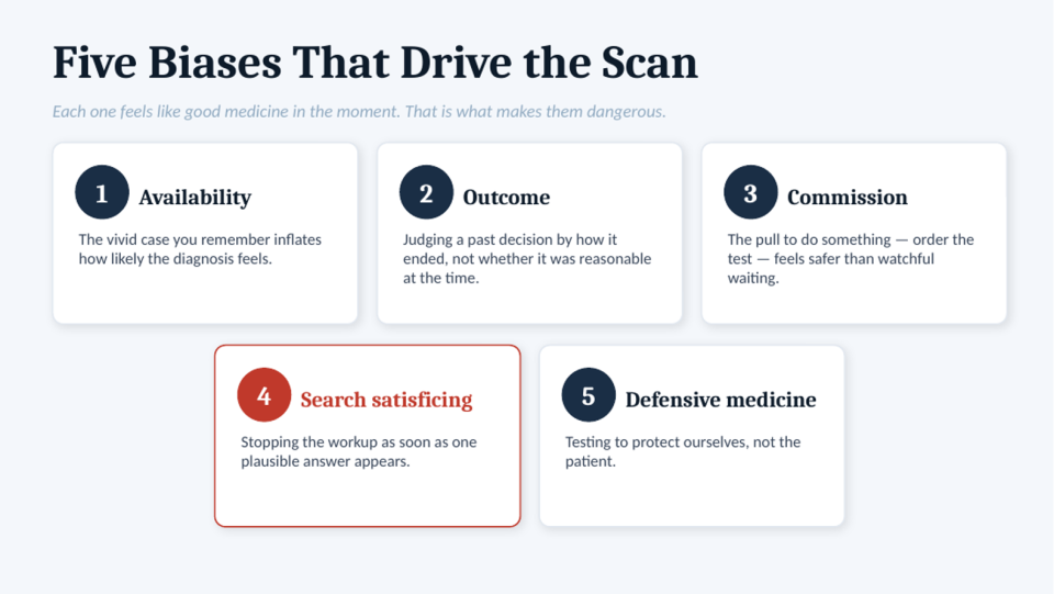
Five biases drive the scan. Each one feels like good medicine in the moment. That is what makes them dangerous.
- Availability bias. The vivid case you remember inflates how likely the diagnosis feels. The plane crash, not the diabetes.
- Outcome bias. Judging a past decision by how it ended, not whether it was reasonable at the time. The missed dissection is remembered for years; the hundreds of times we made the right call and nothing happened are forgotten.
- Commission bias. The pull to do something. Ordering the scan feels active and safe; waiting and watching feels like neglect. But doing something is not automatically safer for the patient. It just feels safer for us.
- Search satisficing. Stopping the workup as soon as one plausible answer appears.
- Defensive medicine. Testing to protect ourselves, not the patient. As the medico-legal data above shows, the fear is often bigger than the actual risk.
Notice the common thread. None of these feels like bad medicine in the moment. We order the scan because we think we are being thorough. We broaden the workup because we think we are being safe. That is what makes them so powerful. They do not feel reckless; they feel responsible. We work in a world where the rare miss is remembered for years while thousands of correct decisions are forgotten by the next day.
Search Satisficing
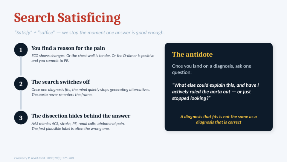
The term combines “satisfy” and “suffice”: we stop looking once we find an answer that seems good enough. For aortic dissection, this is the most dangerous bias of them all. Notice that it works in the opposite direction of the others. Availability bias and commission bias push you toward over-testing. Search satisficing pushes you toward missing the diagnosis altogether.
- You find a reason for the pain. The ECG shows changes, so it is an MI. The chest wall is tender, so it is musculoskeletal. The D-dimer is positive, so you commit to PE.
- The search switches off. Once one diagnosis fits, the mind quietly stops generating alternatives. The aorta never re-enters the frame.
- The dissection hides behind the answer. AAS mimics ACS, stroke, PE, renal colic, and abdominal pain. The first plausible label is often the wrong one, and a diagnosis that fits is not the same as a diagnosis that is correct.
And that is when things go wrong. You diagnose a PE and start heparin, but if the patient actually has a dissection, you have just made things worse. The danger is not only missing the diagnosis. It is treating the wrong one.
The antidote is surprisingly simple. Once you land on a diagnosis, ask one question: “What else could explain this, and have I actively ruled the aorta out, or just stopped looking?”
Dr. Shahbaz Syed (ED Staff at TOH) offers a practical way to think about this: it is fine to form an early impression. In fact, it is efficient. The key is to treat that first diagnosis as a hypothesis, not a conclusion, and then try to disprove it. If you have tried to disprove your diagnosis and it still makes sense, you are probably on the right track.
An answer that fits is not the same as an answer that is right. That single pause to ask “what else” is the highest-value habit we can bring to these patients.
From Reasoning to Practice: The Canadian Guideline
So far, this has all been about how to think. This section is about how to act.
The good news is that you do not need to remember every likelihood ratio, every study, or every percentage discussed above. In 2020, a Canadian clinical practice guideline took all of that complexity and turned it into something practical: one score, three pathways, and one question: how likely is dissection in this patient? Once you answer that, the pathway does the rest. It was developed for Canadian emergency departments by Canadian emergency physicians.
The RIPP Score
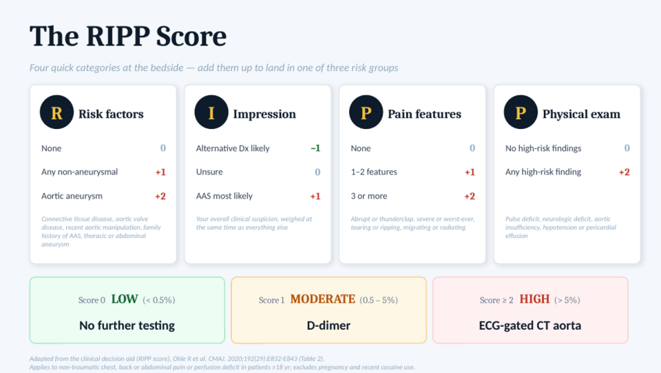
The four letters remind you of the four things to assess: Risk factors, Impression, Pain features, and Physical exam.
- R: Risk factors
- None = 0.
- Any non-aneurysmal risk factor = +1 (connective tissue disease, aortic valve disease, recent aortic manipulation, family history of AAS).
- Aortic aneurysm, thoracic or abdominal, = +2 because it carries the strongest association with dissection.
- I: Impression
- Alternative diagnosis likely = −1.
- Unsure = 0.
- AAS most likely = +1.
This is your overall clinical suspicion, weighed at the same time as everything else. Notably, clinical suspicion ranked higher than any individual risk factor or pain feature.
- P: Pain features
- None = 0.
- One to two features = +1.
- Three or more = +2.
The high-risk features are abrupt or thunderclap onset, severe or worst-ever pain, tearing or ripping quality, and migrating or radiating pain.
- P: Physical exam
- No high-risk findings = 0.
- Any high-risk finding = +2: pulse deficit, neurologic deficit, aortic insufficiency, hypotension, or pericardial effusion.
Then you add everything up:
- Score 0: LOW risk (<0.5%): no further testing.
- Score 1: MODERATE risk (0.5–5%): D-dimer.
- Score ≥2: HIGH risk (>5%): ECG-gated CT aorta.
Does the Score Hold Up?
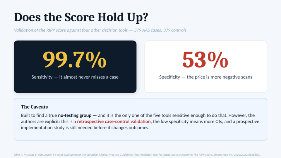
The same group validated the score in 2023, comparing it against four other decision tools using 379 confirmed cases and an equal number of controls.
The headline is sensitivity: 99.7%. In their data, a low-risk score almost never missed a case. That is exactly what you want from a tool designed to tell you when it is safe not to scan.
The trade-off is specificity: 53%. The score will still send some patients to CT who do not have the disease. That is the price of high sensitivity: the harder you try not to miss anything, the more false alarms you accept.
The caveats matter, and the authors are explicit about them. The RIPP score was built to find a true no-testing group, and it is the only one of the five tools sensitive enough to do that. But this is a retrospective case-control validation, the low specificity means more CTs, and a prospective implementation study is still needed before it changes outcomes.
So, this is not a tool to follow blindly. Think of it as a structured way to organize the same reasoning we have been building throughout.
The One-Page Approach
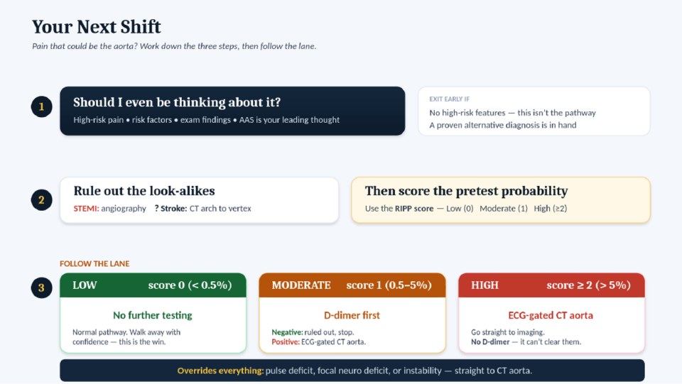
If you take one thing from this to your next shift, make it this. It comes down to three steps.
Step one: should I even be thinking about dissection? This is the step we most often skip. Think about PE for a moment. When a patient tells you their pain is pleuritic, you do not automatically reach for a Wells score and start working up a clot. You first decide whether the overall picture fits. Dissection is no different. Just because the diagnosis crosses your mind does not mean you need to start the pathway. If there are high-risk pain features, risk factors, concerning exam findings, or dissection is genuinely on your differential, continue. If not, stop there. No workup needed.
Step two: do not get fooled by the mimics. Dissection can look like a STEMI, and it can look like a stroke. A STEMI goes to angiography, but remember that dissection can coexist with STEMI. A possible stroke should be scanned from the arch to the vertex so you do not miss the dissection on the way.
Then calculate the RIPP score.
Step three: follow the lane.
- Low risk: no further testing.
- Moderate risk: D-dimer first; if negative, you stop; if positive, go to CT.
- High risk: they have bought themselves a CT.
And one override. If the patient has a pulse deficit, a focal neurologic deficit, or is unstable, bypass the pathway entirely. No score. No D-dimer. Just scan them.
What to Take Home
- The fear is real but miscalibrated. Dissection is genuinely lethal, but it is also extremely uncommon: about one in a thousand chest pain patients.
- Pre-test probability decides everything. The same test result can mean completely different things in different patients.
- Most of our exam findings barely move the needle. The ones that do are pulse deficit, focal neurologic deficit, and hypotension.
- Name the bias to catch it. These biases quietly pull us away from good decision-making.
- Score it, follow the lane, respect the override. The Canadian guideline is there to support your clinical judgment, not replace it.
The hope is that the next time you meet that three-in-the-morning chest pain, the aorta will not make you do it. You will be the judge. And you will decide.
References
- Alter SM, Eskin B, Allegra JR. Diagnosis of Aortic Dissection in Emergency Department Patients is Rare. West J Emerg Med. 2015;16(5):629-631. doi:10.5811/westjem.2015.6.25752
- Lefevre-Scelles A, Jeanmaire P, Freund Y, Joly LM, Phillipon AL, Roussel M. Investigation of pulmonary embolism in patients with chest pain in the emergency department: a retrospective multicenter study. Eur J Emerg Med. 2020;27(5):357-361. doi:10.1097/MEJ.0000000000000680
- Gulati M, Levy PD, Mukherjee D, et al. 2021 AHA/ACC/ASE/CHEST/SAEM/SCCT/SCMR Guideline for the Evaluation and Diagnosis of Chest Pain: A Report of the American College of Cardiology/American Heart Association Joint Committee on Clinical Practice Guidelines. Circulation. 2021;144(22):e368-e454. doi:10.1161/CIR.0000000000001029
- Mandrekar S, Venkatesan P, Nagaraja R. Prevalence of musculoskeletal chest pain in the emergency department: a systematic review and meta-analysis. Scand J Pain. 2021;21(3):434-444. Published 2021 Apr 12. doi:10.1515/sjpain-2020-0168
- Fortier JH, Mostafapour M, Ohle R, et al. Aortic dissection in the ED: a medico-legal perspective on diagnostic delays and failures. CJEM. 2025;27(11):923-931. doi:10.1007/s43678-025-00999-y
- Ohle R, Savage DW, McIsaac S, Yadav K, Caswell J, Conlon M. Epidemiology, mortality and miss rate of acute aortic syndrome in Ontario, Canada: a population-based study. CJEM. 2023;25(1):57-64. doi:10.1007/s43678-022-00413-x
- Ohle R, McIsaac S, Yan J, Yadav K, Eagles D, Perry JJ. National survey of emergency physicians on the risk stratification and acceptable miss rate of acute aortic syndrome. CJEM. 2020;22(3):309-312. doi:10.1017/cem.2019.489
- Ohle R, Kareemi HK, Wells G, Perry JJ. Clinical Examination for Acute Aortic Dissection: A Systematic Review and Meta-analysis. Acad Emerg Med. 2018;25(4):397-412. doi:10.1111/acem.13360
- Klompas M. Does this patient have an acute thoracic aortic dissection?. JAMA. 2002;287(17):2262-2272. doi:10.1001/jama.287.17.2262
- Gaul C, Dietrich W, Erbguth FJ. Neurological symptoms in aortic dissection: a challenge for neurologists. Cerebrovasc Dis. 2008;26(1):1-8. doi:10.1159/000135646
- Um SW, Ohle R, Perry JJ. Bilateral blood pressure differential as a clinical marker for acute aortic dissection in the emergency department. Emerg Med J. 2018;35(9):556-558. doi:10.1136/emermed-2018-207499
- Rogers AM, Hermann LK, Booher AM, et al. Sensitivity of the aortic dissection detection risk score, a novel guideline-based tool for identification of acute aortic dissection at initial presentation: results from the international registry of acute aortic dissection. Circulation. 2011;123(20):2213-2218. doi:10.1161/CIRCULATIONAHA.110.988568
- Ohle R, Anjum O, Bleeker H, McIsaac S. What Is the Specificity of the Aortic Dissection Detection Risk Score in a Low-prevalence Population?. Acad Emerg Med. 2019;26(6):632-638. doi:10.1111/acem.13634
- Nazerian P, Mueller C, Soeiro AM, et al. Diagnostic Accuracy of the Aortic Dissection Detection Risk Score Plus D-Dimer for Acute Aortic Syndromes: The ADvISED Prospective Multicenter Study. Circulation. 2018;137(3):250-258. doi:10.1161/CIRCULATIONAHA.117.029457
- Ren S, Essat M, Pandor A, et al. Diagnostic accuracy of the aortic dissection detection risk score alone or with D-dimer for acute aortic syndromes: Systematic review and meta-analysis. PLoS One. 2024;19(6):e0304401. Published 2024 Jun 21. doi:10.1371/journal.pone.0304401
- Essat M, Goodacre S, Pandor A, Ren S, Ren S, Clowes M. Diagnostic Accuracy of D-Dimer for Acute Aortic Syndromes: Systematic Review and Meta-Analysis. Ann Emerg Med. 2024;84(4):409-421. doi:10.1016/j.annemergmed.2024.05.001
- Croskerry P. The importance of cognitive errors in diagnosis and strategies to minimize them. Acad Med. 2003;78(8):775-780. doi:10.1097/00001888-200308000-00003
- Ohle R, Yan JW, Yadav K, et al. Diagnosing acute aortic syndrome: a Canadian clinical practice guideline. CMAJ. 2020;192(29):E832-E843. doi:10.1503/cmaj.200021
- Ohle R, McIsaac S, Van Drusen M, et al. Evaluation of the Canadian Clinical Practice Guidelines Risk Prediction Tool for Acute Aortic Syndrome: The RIPP Score. Emerg Med Int. 2023;2023:6636800. Published 2023 May 25. doi:10.1155/2023/6636800