
Skin conditions affect approximately one-third of the world’s population and are the 4th most common cause of disease.1,2. As the largest organ in the human body, the skin frequently manifests early signs of various diseases, serving as a pivotal clue in the diagnostic process. Think of Lyme disease as an example: when we recognize the classic rash of erythema migrans and treat early, the systemic complications of the disease can be avoided, but if not recognized early enough, devastating consequences of disease progression can arise.
Dermatology in Skin of Colour: History and Overview
By 2042, more than 50% of the US population is projected to have skin of color. 3 In Ottawa, 26% of residents are people with skin of colour. 4 By 2041, 1 in 4 Canadians will be born in Asia or Africa.5 Given these statistics and future projections, it is imperative that we increase our commitment to providing inclusive care in the Emergency Department.

https://giwps.georgetown.edu/dei-landing-page/
Dermatology as a clinical speciality emerged in Europe in the 19th & 20th centuries.6 Historically skin conditions were described in patients with lighter skin.7 Dermatology relies on pattern recognition, and during our medical training, we are mostly trained to diagnose rashes on lighter skin complexions. Given the differences in how rashes present in skin of colour, this means patients with dark complexion are often undiagnosed or misdiagnosed resulting in a delay to receiving appropriate treatment.
In 2013 a survey among 78 dermatology trainees and junior consultants in the UK showed that only 49% of them thought they would become competent in ethnic dermatology.8 In A US survey, 47% of dermatologists and dermatology residents reported that their training was inadequate in skin conditions in Black patients.9 A survey among 56 dermatologists in Ireland showed that 46% didn’t feel confident diagnosing skin conditions in patients with skin of colour.10 Can you imagine how the average emergency medicine consultant feels about diagnosing rashes in patients with skin of colour?
Fitzpatrick Scale
How does a dermatologist describe different skin tones?
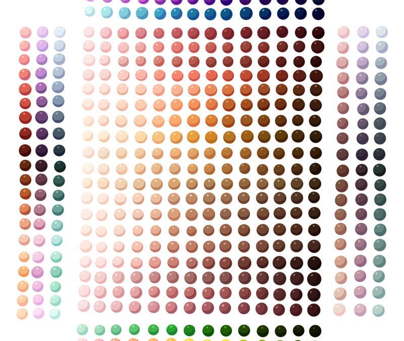
In 1975 a tool called the Fitzpatrick scale was developed the level of tanning vs. burning of an individual’s skin response to UV exposure. The scale has 6 different types as you see here. This is the language that dermatologists use to describe different skin tones.

https://en.wikipedia.org/wiki/Fitzpatrick_scale#/media/File:Influence_of_pigmentation_on_skin_cancer_risk.png
Common Skin Conditions
Unfortunately, there is a paucity of Creative Common Licensed images available online for rashes in skin of colour (ironically, underpins the message of this post). So there are limited images available, the author of this grand rounds specifically refers to VisualDx but those images are unable to be shared here. Some hyperlinks or images contain links to commercially available images to be better help with spot diagnosis.
Eczema (Atopic Dermatitis)
This is a very commonly encountered skin condition in the ED, affecting 36% of pediatric population and up to 23% of the adult population globally. 13,14,15 The erythema that can be appreciated in eczema is seen in patients with skin tones 1-3 as pink or red discolouration contrasted on a light base. What does erythema look like in darker skin tone?
The increased blood flow of erythema does not always appear red or pink in dark skin. If you look only for redness, you may miss the diagnosis of eczema or underestimate disease severity. The red and scaly patches characteristic of eczema in lighter skin tones can sometimes present as lesions that have a grayish, purplish, or deep brown colouration in darker skin tones. Lichenified areas typically appear hyper-pigmented. You can ask the patient if they noticed any change in their skin colour. In darker skin, erythema may be subtle and often overlooked. Sometimes gentle pressure to blanch areas of involvement may help reveal subtle erythema. It is important to recognise erythema in darker skin so that the severity of the eczema is not underestimated. In eczema, dry skin may have a whitish or ash coloured appearance. Post-inflammatory areas are thickened and typically appear hyperpigmented.
Follicular prominence is a characteristic presentation of eczema in patients of African descent. It is rarely seen in types 1-3 skin. In darker skin colours, erythema may be subtle, and papules may appear purple or brown.

Image from https://nationaleczema.org/blog/ate-dark-spots/
Pigmentary Changes
Pigmentary changes are more prominent in darker skin tones and include post-inflammatory hyper-, hypo-, and/or depigmentation. These changes can cause social distress to patients. This is why, early recognition and treatment of eczema are crucial to reduce or prevent long-term pigment abnormalities.
Periorbital eczema creates scaly hyperpigmented plaques on the skin of the upper and lower eyelids. Periorbital hyperpigmentation occurs more frequently in eczema patients with darker skin.
Summary of Findings in Skin of Colour:
- Erythema may be subtle, don’t look for redness, instead look for dark/brown, greyish or purple discolouration
- Follicular prominence is a characteristic presentation of eczema in skin of patients of African descent.
- Periorbital eczema is more common
- Post-inflammatory pigmentary changes are more prominent and can cause social distress to patients.
Psoriasis
Psoriasis is a chronic, relapsing inflammatory disease characterized by demarcated erythematous, silvery, scaly plaques. The primary lesion in psoriasis in patients with lighter skin is a red, scaling papule that further develops into a red, scaling plaque with demarcated borders. In dark skin there is less obvious erythema and lesions may appear violaceous or hyperpigmented, with overlying silvery or grey scale. Importantly, Black individuals often have more extensive cutaneous involvement than White individuals.
Psoriasis in skin of colour is frequently misdiagnosed as a fungal infection. Black women may present with more severe scalp psoriasis. There is a diagnostic delay for Black patients with psoriasis likely because of the educational gap in medical training, and delaying such a diagnosis can bring social and psychological distress to the patient.

Image from AAD
https://www.aad.org/public/diseases/psoriasis/treatment/could-have/skin-color
Summary of Findings in Skin of Colour:
- Erythema is subtle
- Plaques may appear pink, purple, grayish, or dark brown
- Post inflammatory hyperpigmentation of the skin is more common, and can be more severe, and persistent
- Black women may present with severe scalp psoriasis that is often misdiagnosed as a fungal infection
Cellulitis
Cellulitis is a common bacterial infection of the skin that present with erythema, pain, warmth, and swelling. We all know the importance of diagnosing cellulitis early to prevent devastating complications. In darker skin erythema can be subtle. Using a bright light may help spot any subtle colour changes. The presence of tenderness and cobble-stoning on PoCUS may provide additional diagnostic information.
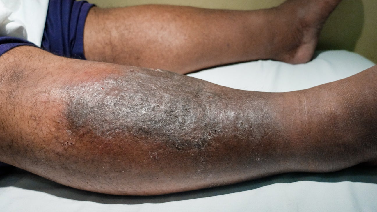
Image from https://www.healthline.com/health/cellulitis
Impetigo
Bullous impetigo is a bacterial skin infection caused by Staph aureus resulting in the formation of large blisters known as bullae. The “classic” presentation that we are taught in medical school describes honey-coloured crusts. In dark skin you do not see honey-coloured crusts and instead crusts may appear gray, purple or dark brown.

Image from https://www.healthline.com/health/impetigo#pictures
Erythema Migrans
Lyme disease is endemic in the Ottawa valley and increasing presenting concern in the Emergency department. We’re trained to recognise the ‘classic rash’ during our medical training; erythema migrans can appear as a hallmark “bull’s-eye” or target-shaped lesion. It is one of the earliest physical exam findings to appreciate in Lyme disease; we know that Lyme disease can lead to devastating systemic complications. That is why early diagnosis and treatment can affect a patient’s morbidity.

https://upload.wikimedia.org/wikipedia/commons/3/34/Adult_deer_tick.jpg
In a study examining patients with Lyme disease, it was observed that Black patients had a greater frequency of arthritis diagnoses (associated with late-stage Lyme disease) and a lower frequency of erythema migrans diagnoses (indicative of early-stage Lyme disease) compared to White patients.16 This may be because erythema migrans presents differently in dark skin and we were not taught on how to recognize it. The contrast of erythema migrans is more readily appreciated in lighter skin colours. In darker skin colours it is more difficult to appreciate erythema; it may appear deep red, purple, or brown.
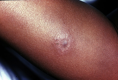
Image from https://www.cdc.gov/lyme/signs_symptoms/rashes.html
“I think the first step for us as ED physicians is to be aware of how EM may present differently in dark skin and not fixate on the classic appearance that we were taught in medical school. When I looked for pictures of this rash in 4 different dermatology textbooks and on the internet, I was only able to find 3 pictures of erythema migrans in patients of colour that’s probably why we are missing the bull’s eye.”
Keloids
Keloids are fibrous tissue nodules usually found on previously traumatized skin or can arise spontaneously. Over weeks to months, these nodules can become painful, pruritic, and expansive; they can cause discomfort, be disfiguring, and restrict normal tissue motion. Although keloids are benign, they are often psychologically and socially devastating for patients, and pose a significant clinical problem for patients of colour; most patients begin developing keloids in their 20s.

https://commons.wikimedia.org/wiki/File:Keloid-Butterfly,_Chest_Wall.JPG
Keloids vs Hypertrophic Scars:
| Keloids | Hypertrophic Scars |
| Invades normal skin | Limited to traumatized area |
| May continue to grow for the patient’s life | Regress spontaneously in 12-18 months |
| Resistant to therapy | Usually responds to therapy |
| Pruritic, painful, burning | Usually asymptomatic |
Keloid Preventative Measures:
- Counsel patients to avoid tattoos, avoid piercings, and avoid elective cosmetic surgery
- Repair skin lacerations with normal tension
Pseudofolliculitis Barbae (PFB)
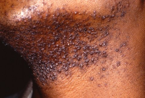
PFB is a common dermatologic disorder of the hair follicles affecting people of colour who shave. The primary lesions of PFB are papules and pustules on the beard area that are disfiguring, with scarring, post inflammatory hyperpigmentation and secondary infection. Perifollicular papules may appear red, purple, gray, or different shades of brown.
https://upload.wikimedia.org/wikipedia/commons/8/83/Pseudofolliculitis_Barbae.jpg
PFB Counselling and Management:
- The only 100% effective preventative treatment is to discontinue shaving
- Counsel patients to to stop shaving and refer to Dermatology
- Once scars or hyperpigmentation develop the skin does not return to normal
Hidradenitis Suppurativa (HS)
HS is a chronic inflammatory condition of the terminal follicular epithelium, which manifests as painful nodules, abscesses and fistulas, most commonly seen on the buttocks, groin breasts and axilla. This condition is more common in people of African descent. HS can be severe with odorous purulent material draining from HS lesions; patients report decreased quality of life and are at an increased risk of suicide.17 In patients with skin tones 1-3, HS usually presents with pustules, pink or red erythematous nodules and linear scarring. In darker skin patients the erythema of papules, nodules and cysts are challenging to see; the lesions may appear dark brown, purple, gray.
HS Management:
- Focus on treating pain, and be aware of the social factors
- Refer to Dermatology
- Refer to mental health support services when indicated
- Lifestyle counselling – weight loss and smoking cessation may reduce the frequency of HS flares
Morbilliform Drug Eruption:
Morbilliform drug eruptions are the most common medication-induced rash. These reactions can vary from exanthematous eruptions to life-threatening conditions with multisystem involvement. In skin tones 1-3 the rash usually presents as red macules and papules that arise on the trunk and spread symmetrically to the proximal extremities, but in patients with darker skin clinical findings may be subtle, presenting as purple or gray and more challenging to diagnose. These maculopapular eruptions are often underappreciated in dark skin.
For patients with darker skin post inflammatory hyper or hypo-pigmentation may be more prominent and may take weeks to months to resolve. As in other erythematous skin conditions it is important to ask the patient if they have noticed any change to the colour of their skin. A complete physical exam should include the external skin surface completely, oral mucosa, genitals, ocular area and lymph nodes.

Image from https://www.ncbi.nlm.nih.gov/pmc/articles/PMC9952815/ 18
Drug Induced Hypersensitivity Syndrome (DIHS)
DIHS, previously known as drug reaction with eosinophilia and systemic symptoms (DRESS), is an idiosyncratic drug reaction with systemic manifestations including rash, fever and internal organ involvement (most typically hepatitis). An exanthematous eruption is usually present, and erythema in dark skin may be subtle and appear gray or purple. If you suspect your patient may have drug induced hypersensitivity syndrome, full examination of the skin and oral mucosa is important and blood work including LFTs to rule out internal organ involvement.
References:
- Giesey RL, Mehrmal S, Uppal P, et al. The global burden of skin and subcutaneous disease: a longitudinal analysis from the Global Burden of Disease Study from 1990-2017. Skin. 2021;5(2):125-136.
- Flohr C, Hay R. Putting the burden of skin diseases on the global map. Br J Dermatol. 2021;184:189-190.)
- Sauaia A, Dellavalle RP. Health care inequities: an introduction for dermatology providers. Dermatol Clin. 2009 Apr;27(2):103-7, v. doi: 10.1016/j.det.2008.12.001. PMID: 19254652; PMCID: PMC2677554.
- https://www12.statcan.gc.ca/census-recensement/2016/dp-pd/prof/index.cfm?Lang=E
- https://www150.statcan.gc.ca/n1/daily-quotidien/220908/dq220908a-eng.htm
- Myers J. Challenges of identifying eczema in darkly pigmented skin. Nurs Child Young People 2015; 27: 24–8. DOI: 10.7748/ncyp.27.6.24.e571.
- Ogunyemi B, Miller-Monthrope Y. The state of ethnic dermatology in Canada. J Cutan Med Surg 2017; 21: 464–6. DOI: 10.1177/1203475417711110
- Salam, O.E. Dadzie, Dermatology training in the U.K.: does it reflect the changing demographics of our population?, British Journal of Dermatology, Volume 169, Issue 6, 1 December 2013, Pages 1360–1362, https://doi.org/10.1111/bjd.12491
- Buster KJ, Stevens EI, Elmets CA. Dermatologic health disparities. Dermatol Clin. 2012 Jan;30(1):53-9, viii. doi: 10.1016/j.det.2011.08.002. PMID: 22117867; PMCID: PMC3742002.
- O’Connor, C. Gallagher, J. Bourke, M. Murphy, Confidence of Irish dermatologists in caring for patients with skin of colour, Clinical and Experimental Dermatology, Volume 47, Issue 1, 1 January 2022, Pages 169–171, https://doi.org/10.1111/ced.14897
- Adelekun A, Onyekaba G, Lipoff JB. Skin color in dermatology textbooks: An updated evaluation and analysis. J Am Acad Dermatol. 2021 Jan;84(1):194-196. doi: 10.1016/j.jaad.2020.04.084. Epub 2020 Apr 23. PMID: 32335181.
- Louie P, Wilkes R. Representations of race and skin tone in medical textbook imagery. Soc Sci Med. 2018 Apr;202:38-42. doi: 10.1016/j.socscimed.2018.02.023. Epub 2018 Feb 23. PMID: 29501717.
- Nutten, S. (2015). Atopic dermatitis: global epidemiology and risk factors. Ann Nutr Metab, 66(suppl 1), 8-16. https://doi.org/10.1159/000370220
- Bylund, S., von Kobyletzki, L.B., Svalstedt, M., Svensson, Å. (2020). Prevalence and Incidence of Atopic Dermatitis: A Systematic Review. Acta Derm Venereol, 100(12). https://www.doi.org/10.2340/00015555-3510
- Mathiesen, S.M., Thomsen, S.F. (2019). The prevalence of atopic dermatitis in adults: systematic review on population studies. Dermatol Online J, 25(8). https://doi.org/10.5070/D3258045124
- Fix AD, Peña CA, Strickland GT. Racial differences in reported Lyme disease incidence. Am J Epidemiol 2000;152:756-759
- Chernyshov PV, Finlay AY, Tomas-Aragones L, Poot F, Sampogna F, Marron SE, Zemskov SV, Abeni D, Tzellos T, Szepietowski JC, Zouboulis CC. Quality of Life in Hidradenitis Suppurativa: An Update. Int J Environ Res Public Health. 2021 Jun 6;18(11):6131. doi: 10.3390/ijerph18116131. PMID: 34204126; PMCID: PMC8201351.
- Lehloenya RJ, Phillips EJ, Pasieka HB, Peter J. Recognizing Drug Hypersensitivity in Pigmented Skin. Immunol Allergy Clin North Am. 2022 May;42(2):219-238. doi: 10.1016/j.iac.2022.01.005. Epub 2022 Mar 31. PMID: 35469616; PMCID: PMC9952815.


This is very useful