Abdominal Aortic Aneurysm
Modality: Still or Cineclip
Minimum Archiving Requirement:
- If no AAA present, transverse images of the proximal, mid, distal and bifurcation of the aorta
- If AAA present, single transverse image
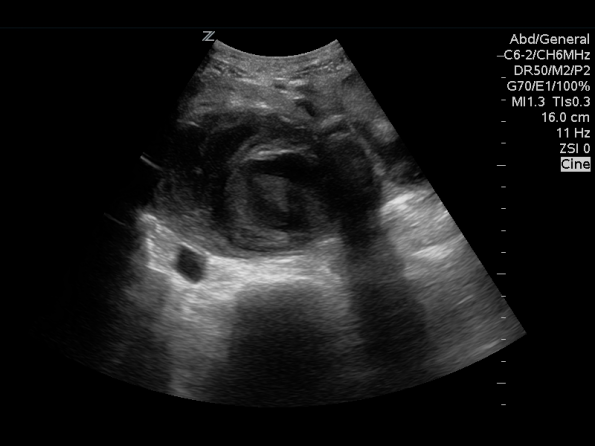
ultrasound
AAA present
No AAA, Proximal Aorta

No AAA, Mid Aorta
No AAA, Aorta Distal Bifurcation
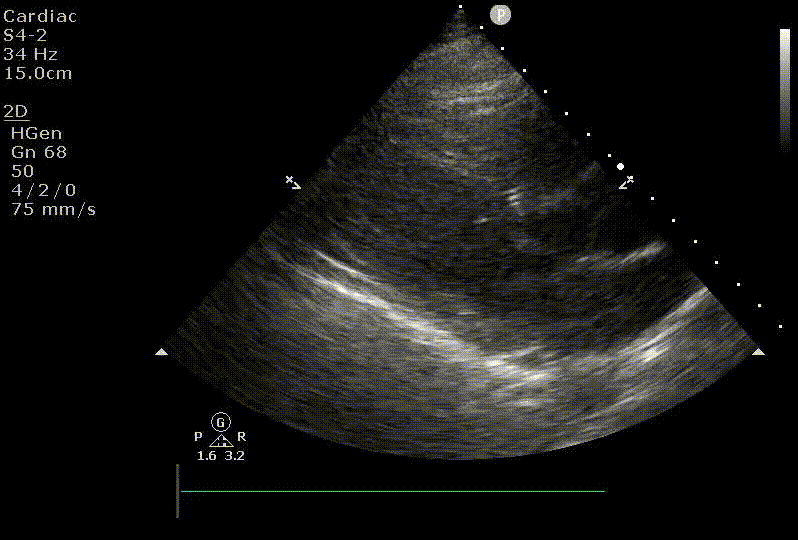
Cardiac Arrest
Modality: Cineclip
Minimum Archiving Requirement: Best possible view
Cardiac Arrest
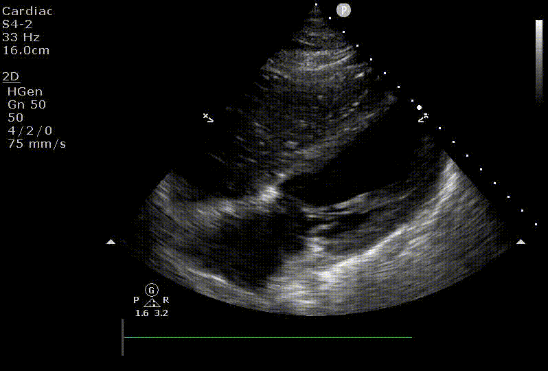
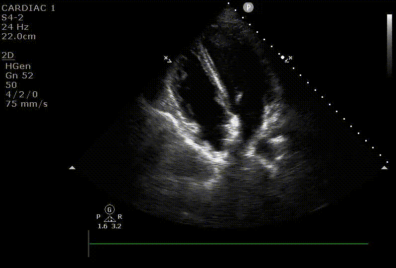
Left Ventricular Function
Modality: Cineclip
Minimum Archiving Requirement: Parasternal long axis view
LV Function
Right Ventricular Function
Modality: Cineclip
Minimum Archiving Requirement: Apical 4-chamber view
RV function
Pericardial Effusion
Modality: Cineclip
Minimum Archiving Requirement: Best possible view, subxiphoid is the most common
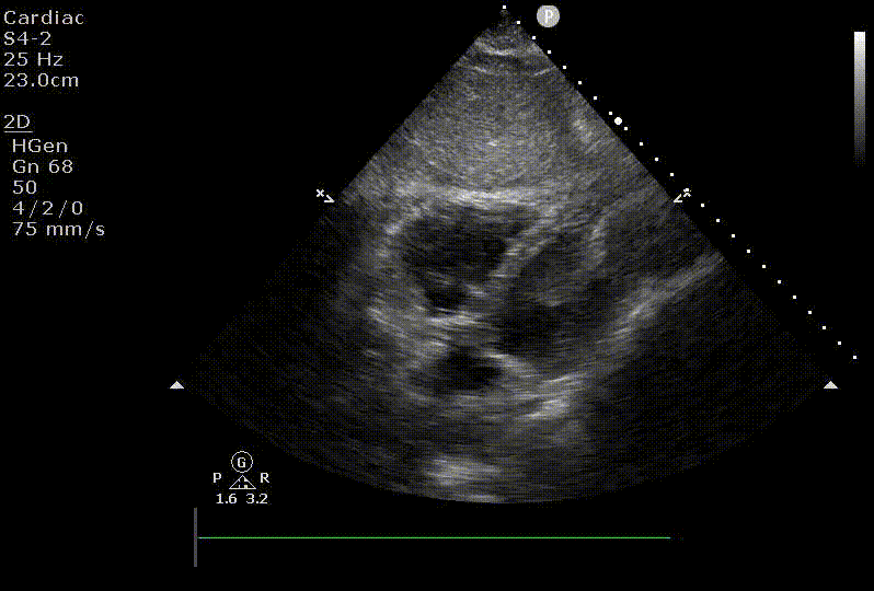
Pericardial Effusion
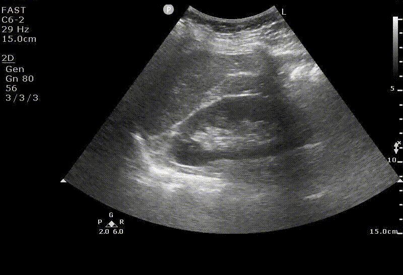
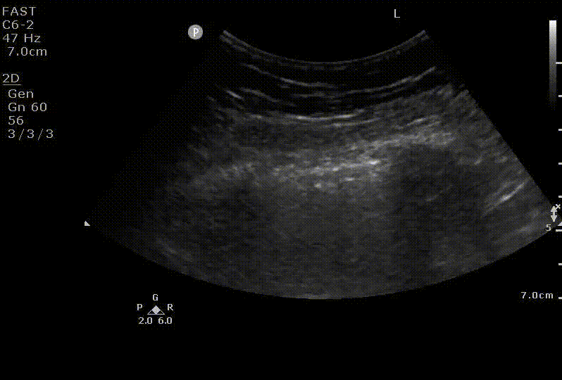
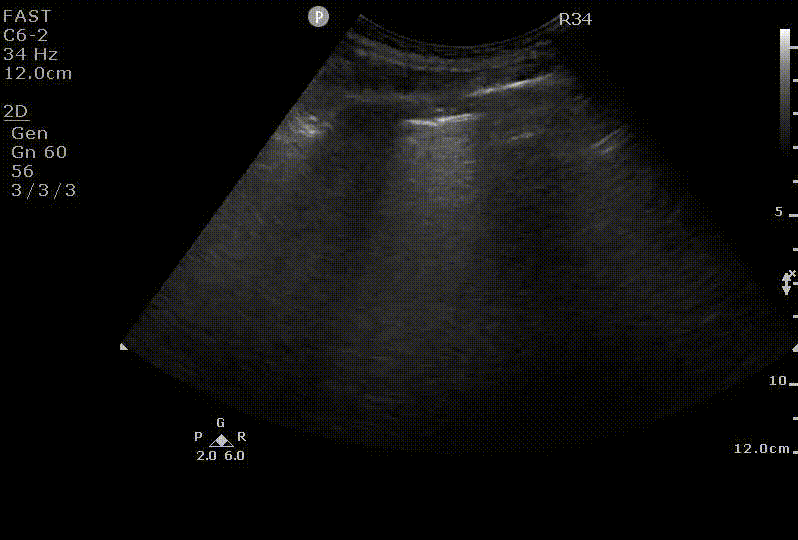
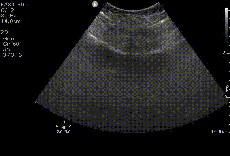
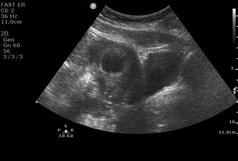
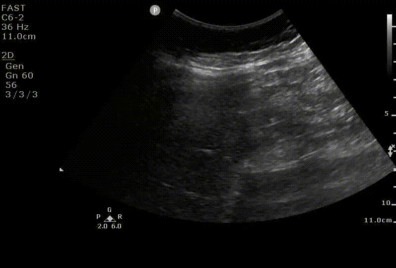
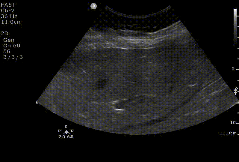
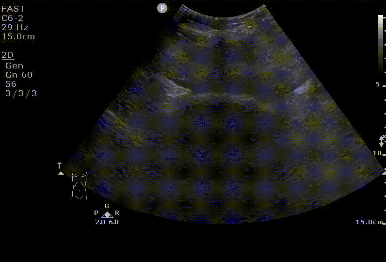
Focused assessment with sonography for trauma (FAST)
Modality: Still or Cineclip
Minimum Archiving Requirement: Parasternal long axis view
- Entire hepatorenal interface to the tip of the liver (Morison’s pouch)
- Diaphragm-liver interface up to the 9 o’clock position (including R pleuraleffusion)
- Entire splenorenal interface including the caudal spleen tip
- Diaphragm-spleen interface up to the 9 o’clock position (including L pleural effusion)
- Transverse view of the pelvis
- Best possible cineclip to assess for pericardial effusion
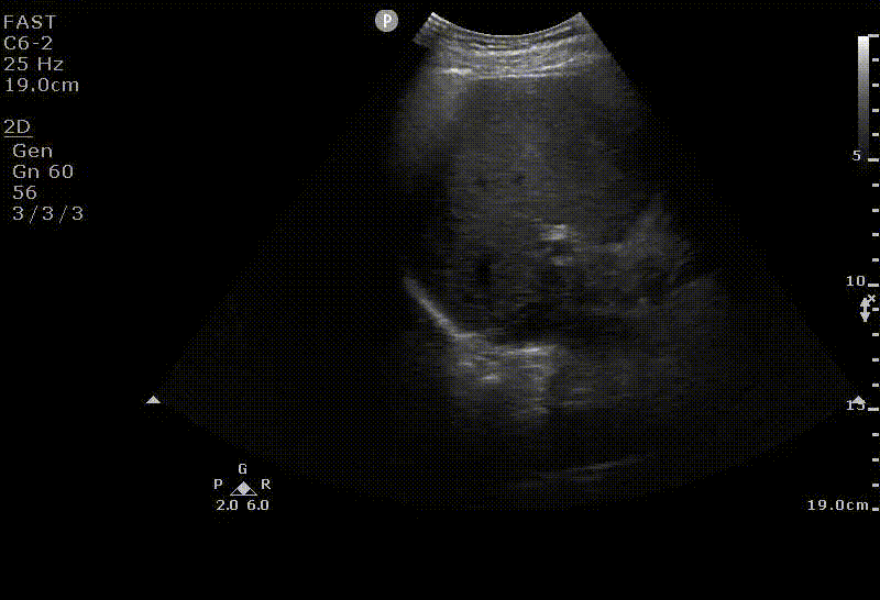
RUQ
LUQ
Pelvis
Pneumothorax
Modality: Cineclip or M-Mode
Minimum Archiving Requirement: Bilateral sagittal scans at the mid-clavicular line, 3rd intercostal space
Left Lung (repeat on contralateral side)
Pulmonary Edema
Modality: Cineclip
Minimum Archiving Requirement: Minimum of 2 representative zones, bilaterally
Pulmonary Edema
Modality: Cineclip
Minimum Archiving Requirement: Minimum of 2 representative zones, bilaterally
Pregnancy of Unknown Location
Modality: Still or Cineclip
Minimum Archiving Requirement:
- Sagittal and transverse views of the uterus and bladder
- Gestational sac in the uterus, if present
- Fetal pole and yolk sac, if present
- Myometrial mantle measurement if it appears abnormal
- Fetal heart rate documentation, if present, using M-mode still or cine-clips
Transverse
Sagittal
Myometrial Mantle
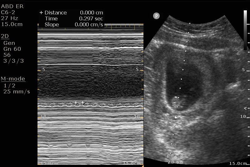
Fetal Heart Rate
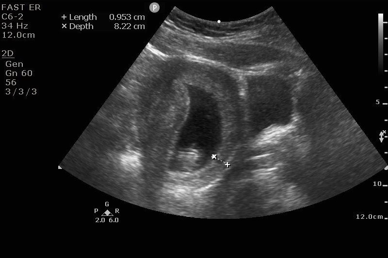
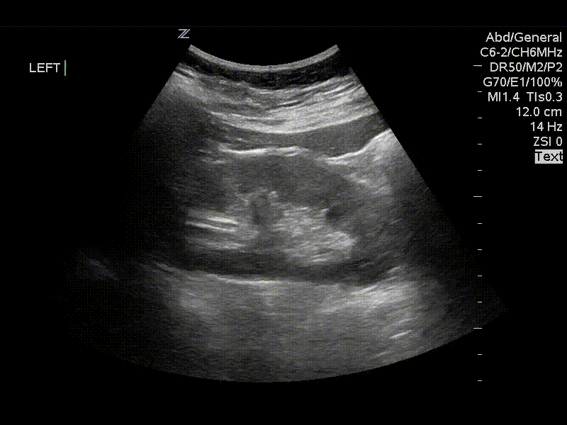
Cholelithiasis and Cholecystitis
Check out our gallbladder PoCUS learning module here
Modality: Still or Cineclip
Minimum Archiving Requirement:
- Long axis view
- Short axis view
- In the context of cholecystitis, measure of the anterior wall of the gallbladder
Short Axis
Long Axis

Long Axis
Cholecystitis Measurement
Hydronephrosis
Modality: Still or Cineclip
Minimum Archiving Requirement:
- Long axis view of bilateral kidneys
- Transverse view of the bladder
Left Kidney
Pelvic
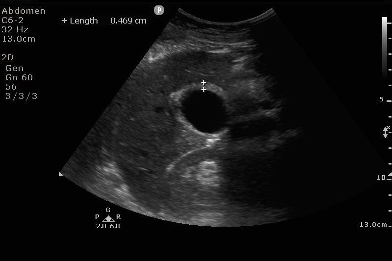
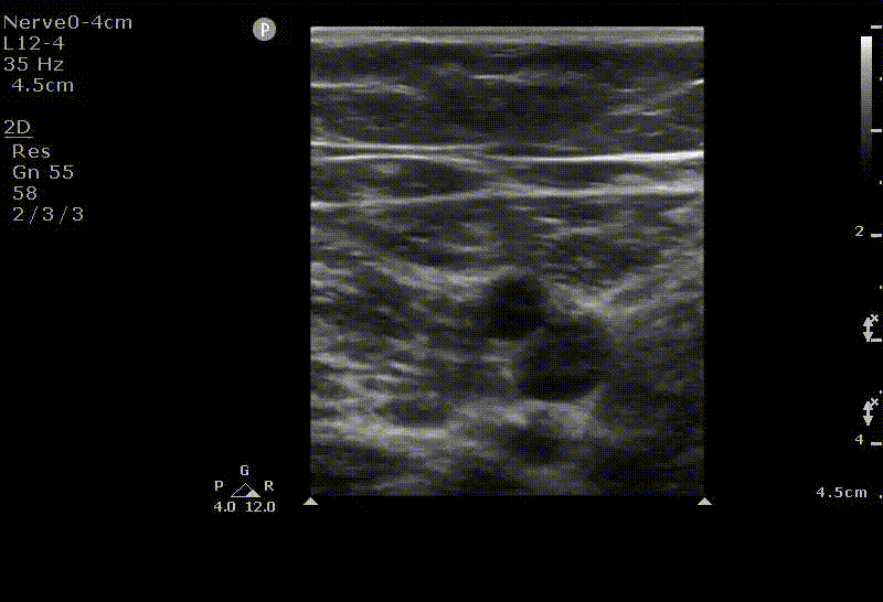
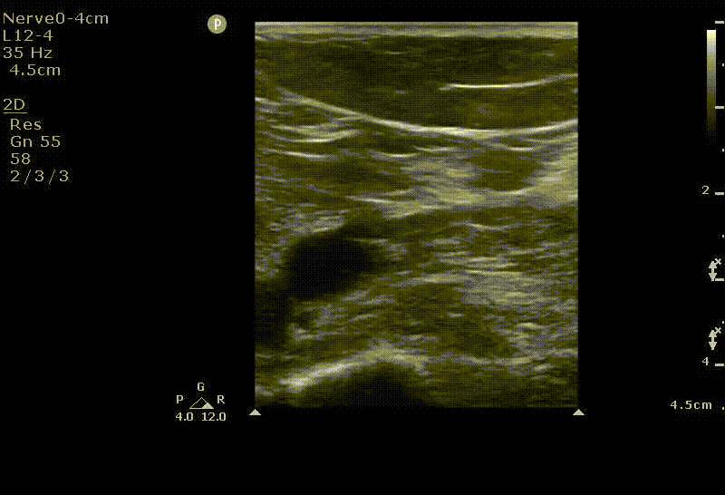
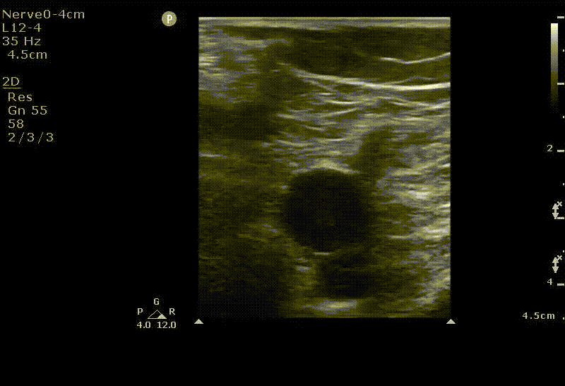
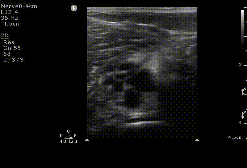
Above knee lower extremity Deep Venous Thrombosis
Modality: Cineclip with and without compression
Minimum Archiving Requirement:
- Zone 1
- Common femoral vein
- Saphenous femoral junction
- Femoral vein beyond the deep femoral branch
- Zone 2
- Distal femoral vein
- Popliteal vein
- Popliteal vein trifurcation
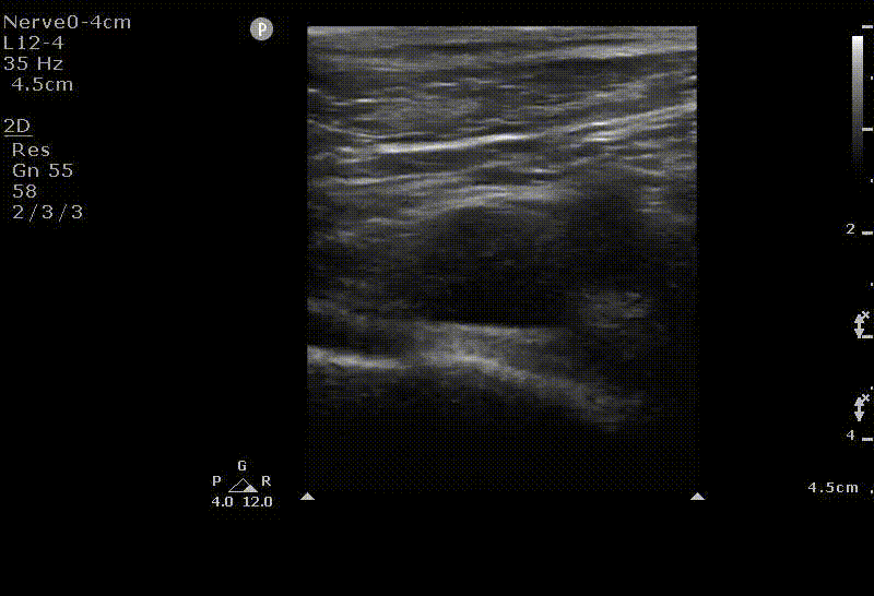
Zone 1: Common Femoral Vein
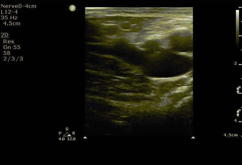
Zone 1: Saphenous Femoral Junction
Zone 1: Femoral Vein
Zone 2: Distal Femoral Vein
Zone 2: Popliteal Vein
Zone 2: Popliteal Zone Trifurcation


Central Line Insertion
Modality: N/A
Minimum Archiving Requirement: No routine archiving necessary; if choosing to archive, a confirmatory image of wire in vein